Crystal Structures of Aspartate Aminotransferase Reconstituted with 1-Deazapyridoxal 5'-Phosphate: Internal Aldimine and Stable l-Aspartate External Aldimine.
Griswold, W.R., Fisher, A.J., Toney, M.D.(2011) Biochemistry 50: 5918-5924
- PubMed: 21627105 Search on PubMed
- DOI: https://doi.org/10.1021/bi200436y
- Primary Citation Related Structures:
3QN6, 3QPG - PubMed Abstract:
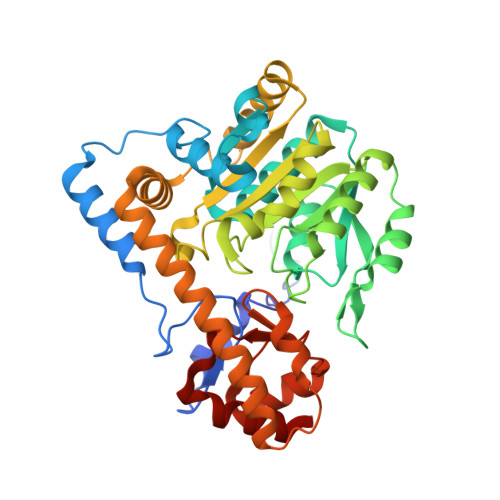
The 1.8 Å resolution crystal structures of Escherichia coli aspartate aminotransferase reconstituted with 1-deazapyridoxal 5'-phosphate (deazaPLP; 2-formyl-3-hydroxy-4-methylbenzyl phosphate) in the internal aldimine and L-aspartate external aldimine forms are reported. The L-aspartate·deazaPLP external aldimine is extraordinarily stable (half-life of >20 days), allowing crystals of this intermediate to be grown by cocrystallization with L-aspartate. This structure is compared to that of the α-methyl-L-aspartate·PLP external aldimine. Overlays with the corresponding pyridoxal 5'-phosphate (PLP) aldimines show very similar orientations of deazaPLP with respect to PLP. The lack of a hydrogen bond between Asp222 and deazaPLP, which serves to "anchor" PLP in the active site, releases strain in the deazaPLP internal aldimine that is enforced in the PLP internal aldimine [Hayashi, H., Mizuguchi, H., Miyahara, I., Islam, M. M., Ikushiro, H., Nakajima, Y., Hirotsu, K., and Kagamiyama, H. (2003) Biochim. Biophys. Acta1647, 103] as evidenced by the planarity of the pyridine ring and the Schiff base linkage with Lys258. Additionally, loss of this anchor causes a 10° greater tilt of deazaPLP toward the substrate in the external aldimine. An important mechanistic difference between the L-aspartate·deazaPLP and α-methyl-L-aspartate·PLP external aldimines is a hydrogen bond between Gly38 and Lys258 in the former, positioning the catalytic base above and approximately equidistant between Cα and C4'. In contrast, in the α-methyl-L-aspartate·PLP external aldimine, the ε-amino group of Lys258 is rotated ~70° to form a hydrogen bond to Tyr70 because of the steric bulk of the methyl group.
- Department of Chemistry, University of California, Davis, California 95616, United States.
Organizational Affiliation: