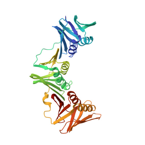
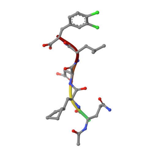
3Q4L

Structure of a small peptide ligand bound to E.coli DNA sliding clamp


Experimental Data Snapshot

  • Method: X-RAY DIFFRACTION
  • Resolution: 1.95 Å
  • R-Value Free: 0.232 
  • R-Value Work: 0.199 
  • R-Value Observed: 0.201 

wwPDB Validation   3D Report Full Report


This is version 1.2 of the entry. See complete history


Literature

Structure-based design of short peptide ligands binding onto the E. coli processivity ring.

Wolff, P.Olieric, V.Briand, J.P.Chaloin, O.Dejaegere, A.Dumas, P.Ennifar, E.Guichard, G.Wagner, J.Burnouf, D.Y.

(2011) J Med Chem 54: 4627-4637

  • DOI: https://doi.org/10.1021/jm200311m
  • Primary Citation of Related Structures:  
    3Q4J, 3Q4K, 3Q4L

  • PubMed Abstract: 

    The multimeric DNA sliding clamps confer high processivity to replicative DNA polymerases and are also binding platforms for various enzymes involved in DNA metabolism. These enzymes interact with the clamp through a small peptide that binds into a hydrophobic pocket which is a potential target for the development of new antibacterial compounds. Starting from a generic heptapeptide, we used a structure-based strategy to improve the design of new peptide ligands. Chemical modifications at specific residues result in a dramatic increase of the interaction as measured by SPR and ITC. The affinity of our best hits was improved by 2 orders of magnitude as compared to the natural ligand, reaching 10(-8) M range. The molecular basis of the interactions was analyzed by solving the co-crystal structures of the most relevant peptides bound to the clamp and reveals how chemical modifications establish new contacts and contributes to an increased affinity of the ligand.


  • Organizational Affiliation

    Architecture et Réactivité de l'ARN, Université de Strasbourg, Institut de Biologie Moléculaire et Cellulaire, Strasbourg, France.


Macromolecules
Find similar proteins by:  (by identity cutoff)  |  3D Structure
Entity ID: 1
MoleculeChains Sequence LengthOrganismDetailsImage
DNA polymerase III subunit beta
A, B
368Escherichia coli K-12Mutation(s): 0 
Gene Names: dnaNb3701JW3678
EC: 2.7.7.7
UniProt
Find proteins for P0A988 (Escherichia coli (strain K12))
Explore P0A988 
Go to UniProtKB:  P0A988
Entity Groups  
Sequence Clusters30% Identity50% Identity70% Identity90% Identity95% Identity100% Identity
UniProt GroupP0A988
Sequence Annotations
Expand
  • Reference Sequence

Find similar proteins by:  Sequence   |   3D Structure  

Entity ID: 2
MoleculeChains Sequence LengthOrganismDetailsImage
peptide ligand
C, D
6N/AMutation(s): 0 
Sequence Annotations
Expand
  • Reference Sequence
Small Molecules
Ligands 1 Unique
IDChains Name / Formula / InChI Key2D Diagram3D Interactions
NA
Query on NA

Download Ideal Coordinates CCD File 
E [auth B]SODIUM ION
Na
FKNQFGJONOIPTF-UHFFFAOYSA-N
Modified Residues  2 Unique
IDChains TypeFormula2D DiagramParent
ALC
Query on ALC
C, D
L-PEPTIDE LINKINGC9 H17 N O2ALA
ZCL
Query on ZCL
C, D
L-PEPTIDE LINKINGC9 H9 Cl2 N O2PHE
Biologically Interesting Molecules (External Reference) 1 Unique
Experimental Data & Validation

Experimental Data

  • Method: X-RAY DIFFRACTION
  • Resolution: 1.95 Å
  • R-Value Free: 0.232 
  • R-Value Work: 0.199 
  • R-Value Observed: 0.201 
  • Space Group: P 1
Unit Cell:
Length ( Å )Angle ( ˚ )
a = 34.84α = 65.28
b = 79.57β = 75.26
c = 81.64γ = 82.22
Software Package:
Software NamePurpose
XSCALEdata processing
BUSTER-TNTrefinement
PDB_EXTRACTdata extraction
XDSdata reduction
XSCALEdata scaling
MOLREPphasing
BUSTERrefinement

Structure Validation

View Full Validation Report


Entry History 

Deposition Data

Revision History  (Full details and data files)

  • Version 1.0: 2011-12-28
    Type: Initial release
  • Version 1.1: 2012-12-12
    Changes: Other
  • Version 1.2: 2013-03-20
    Changes: Database references