3PL6

Structure of Autoimmune TCR Hy.1B11 in complex with HLA-DQ1 and MBP 85-99


Experimental Data Snapshot

  • Method: X-RAY DIFFRACTION
  • Resolution: 2.55 Å
  • R-Value Free: 0.258 
  • R-Value Work: 0.232 

wwPDB Validation   3D Report Full Report


Ligand Structure Quality Assessment 


This is version 1.5 of the entry. See complete history


Literature

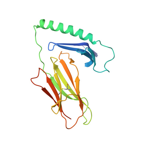
A highly tilted binding mode by a self-reactive T cell receptor results in altered engagement of peptide and MHC.

Sethi, D.K.Schubert, D.A.Anders, A.K.Heroux, A.Bonsor, D.A.Thomas, C.P.Sundberg, E.J.Pyrdol, J.Wucherpfennig, K.W.

(2011) J Exp Med 208: 91-102

  • DOI: https://doi.org/10.1084/jem.20100725
  • Primary Citation of Related Structures:  
    3PL6

  • PubMed Abstract: 

    Self-reactive T cells that escape elimination in the thymus can cause autoimmune pathology, and it is therefore important to understand the structural mechanisms of self-antigen recognition. We report the crystal structure of a T cell receptor (TCR) from a patient with relapsing-remitting multiple sclerosis that engages its self-peptide-major histocompatibility complex (pMHC) ligand in an unusual manner. The TCR is bound in a highly tilted orientation that prevents interaction of the TCR-α chain with the MHC class II β chain helix. In this structure, only a single germline-encoded TCR loop engages the MHC protein, whereas in most other TCR-pMHC structures all four germline-encoded TCR loops bind to the MHC helices. The tilted binding mode also prevents peptide contacts by the short complementarity-determining region (CDR) 3β loop, and interactions that contribute to peptide side chain specificity are focused on the CDR3α loop. This structure is the first example in which only a single germline-encoded TCR loop contacts the MHC helices. Furthermore, the reduced interaction surface with the peptide may facilitate TCR cross-reactivity. The structural alterations in the trimolecular complex are distinct from previously characterized self-reactive TCRs, indicating that there are multiple unusual ways for self-reactive TCRs to bind their pMHC ligand.


  • Organizational Affiliation

    Department of Cancer Immunology and AIDS, Dana-Farber Cancer Institute, Boston, MA 02115, USA.


Macromolecules
Find similar proteins by:  (by identity cutoff)  |  3D Structure
Entity ID: 1
MoleculeChains Sequence LengthOrganismDetailsImage
MHC class II HLA-DQ-alpha chain194Homo sapiensMutation(s): 0 
Gene Names: HLA-DQA1
UniProt
Find proteins for A0A173ADG5 (Homo sapiens)
Explore A0A173ADG5 
Go to UniProtKB:  A0A173ADG5
Entity Groups  
Sequence Clusters30% Identity50% Identity70% Identity90% Identity95% Identity100% Identity
UniProt GroupA0A173ADG5
Sequence Annotations
Expand
  • Reference Sequence
Find similar proteins by:  (by identity cutoff)  |  3D Structure
Entity ID: 2
MoleculeChains Sequence LengthOrganismDetailsImage
MHC class II HLA-DQ-beta chain202Homo sapiensMutation(s): 0 
Gene Names: HLA-DQB1
UniProt
Find proteins for A0A0A8WFP6 (Homo sapiens)
Explore A0A0A8WFP6 
Go to UniProtKB:  A0A0A8WFP6
Entity Groups  
Sequence Clusters30% Identity50% Identity70% Identity90% Identity95% Identity100% Identity
UniProt GroupA0A0A8WFP6
Sequence Annotations
Expand
  • Reference Sequence
Find similar proteins by:  (by identity cutoff)  |  3D Structure
Entity ID: 3
MoleculeChains Sequence LengthOrganismDetailsImage
T-cell receptor alpha chain206Homo sapiensMutation(s): 0 
Entity Groups  
Sequence Clusters30% Identity50% Identity70% Identity90% Identity95% Identity100% Identity
Sequence Annotations
Expand
  • Reference Sequence
Find similar proteins by:  (by identity cutoff)  |  3D Structure
Entity ID: 4
MoleculeChains Sequence LengthOrganismDetailsImage
MBP peptide / T-cell receptor beta chain chimera268Homo sapiensMutation(s): 0 
Entity Groups  
Sequence Clusters30% Identity50% Identity70% Identity90% Identity95% Identity100% Identity
Sequence Annotations
Expand
  • Reference Sequence
Small Molecules
Ligands 1 Unique
IDChains Name / Formula / InChI Key2D Diagram3D Interactions
NAG
Query on NAG

Download Ideal Coordinates CCD File 
E [auth A],
F [auth B]
2-acetamido-2-deoxy-beta-D-glucopyranose
C8 H15 N O6
OVRNDRQMDRJTHS-FMDGEEDCSA-N
Experimental Data & Validation

Experimental Data

  • Method: X-RAY DIFFRACTION
  • Resolution: 2.55 Å
  • R-Value Free: 0.258 
  • R-Value Work: 0.232 
  • Space Group: P 21 21 21
Unit Cell:
Length ( Å )Angle ( ˚ )
a = 73.123α = 90
b = 124.815β = 90
c = 134.921γ = 90
Software Package:
Software NamePurpose
CNSrefinement
PDB_EXTRACTdata extraction
CBASSdata collection
HKL-2000data reduction
HKL-2000data scaling
PHASERphasing

Structure Validation

View Full Validation Report


Ligand Structure Quality Assessment 


Entry History 

Deposition Data

Revision History  (Full details and data files)

  • Version 1.0: 2010-12-22
    Type: Initial release
  • Version 1.1: 2011-07-13
    Changes: Version format compliance
  • Version 1.2: 2017-07-26
    Changes: Source and taxonomy
  • Version 1.3: 2017-11-08
    Changes: Refinement description
  • Version 1.4: 2020-07-29
    Type: Remediation
    Reason: Carbohydrate remediation
    Changes: Data collection, Database references, Derived calculations, Structure summary
  • Version 1.5: 2024-03-13
    Changes: Data collection, Database references, Source and taxonomy, Structure summary