3PIC

Glucuronoyl Esterase catalytic domain (Cip2_GE) from Hypocrea jecorina


Experimental Data Snapshot

  • Method: X-RAY DIFFRACTION
  • Resolution: 1.90 Å
  • R-Value Free: 0.234 
  • R-Value Work: 0.195 
  • R-Value Observed: 0.195 

wwPDB Validation   3D Report Full Report


Ligand Structure Quality Assessment 


This is version 1.3 of the entry. See complete history


Literature

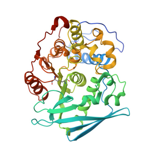
Structure of the catalytic domain of glucuronoyl esterase Cip2 from Hypocrea jecorina.

Pokkuluri, P.R.Duke, N.E.Wood, S.J.Cotta, M.A.Li, X.L.Biely, P.Schiffer, M.

(2011) Proteins 79: 2588-2592

  • DOI: https://doi.org/10.1002/prot.23088
  • Primary Citation of Related Structures:  
    3PIC

  • PubMed Abstract: 

    The structure of the catalytic domain of glucuronoyl esterase Cip2 from the fungus H. jecorina was determined at a resolution of 1.9 Å. This is the first structure of the newly established carbohydrate esterase family 15. The structure has revealed the residues Ser278-His411-Glu301 present in a triad arrangement as the active site. Ser278 is present in the novel consensus sequence GCSRXG reported earlier in the members of CE-15 family. The active site is exposed on the surface of the protein which has implications for the ability of the enzyme to hydrolyze ester bonds of large substrates. Efforts are underway to obtain crystals of Cip2_GE complexed with inhibitor and synthetic substrates. The activity of the glucuronoyl esterase could play a significant role in plant biomass degradation as its expected role is to separate the lignin from hemicelluloses by hydrolysis of the ester bond between 4-O-methyl-D-glucuronic acid moieties of glucuronoxylans and aromatic alcohols of lignin.


  • Organizational Affiliation

    Biosciences Division, Argonne National Laboratory, Argonne, Illinois 60439, USA. rajp@anl.gov


Macromolecules
Find similar proteins by:  (by identity cutoff)  |  3D Structure
Entity ID: 1
MoleculeChains Sequence LengthOrganismDetailsImage
Cip2
A, B, C
375Trichoderma reeseiMutation(s): 0 
UniProt
Find proteins for G0RV93 (Hypocrea jecorina (strain QM6a))
Explore G0RV93 
Go to UniProtKB:  G0RV93
Entity Groups  
Sequence Clusters30% Identity50% Identity70% Identity90% Identity95% Identity100% Identity
UniProt GroupG0RV93
Sequence Annotations
Expand
  • Reference Sequence
Experimental Data & Validation

Experimental Data

  • Method: X-RAY DIFFRACTION
  • Resolution: 1.90 Å
  • R-Value Free: 0.234 
  • R-Value Work: 0.195 
  • R-Value Observed: 0.195 
  • Space Group: P 21 21 21
Unit Cell:
Length ( Å )Angle ( ˚ )
a = 80.26α = 90
b = 82.01β = 90
c = 185.89γ = 90
Software Package:
Software NamePurpose
HKL-2000data collection
HKL-3000phasing
CNSrefinement
HKL-2000data reduction
HKL-2000data scaling

Structure Validation

View Full Validation Report


Ligand Structure Quality Assessment 


Entry History 

Deposition Data

Revision History  (Full details and data files)

  • Version 1.0: 2011-06-01
    Type: Initial release
  • Version 1.1: 2011-07-13
    Changes: Version format compliance
  • Version 1.2: 2011-07-20
    Changes: Database references
  • Version 1.3: 2020-07-29
    Type: Remediation
    Reason: Carbohydrate remediation
    Changes: Data collection, Database references, Derived calculations, Structure summary