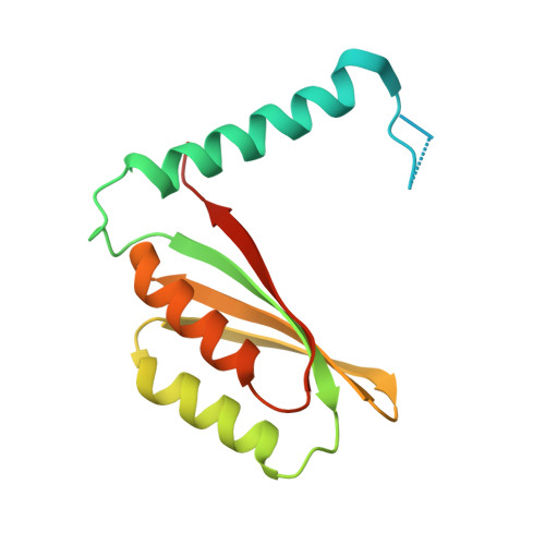
Ni(II) coordination to mixed sites modulates DNA binding of HpNikR via a long-range effect.
West, A.L., Evans, S.E., Gonzalez, J.M., Carter, L.G., Tsuruta, H., Pozharski, E., Michel, S.L.(2012) Proc Natl Acad Sci U S A 109: 5633-5638
- PubMed: 22451934 Search on PubMedSearch on PubMed Central
- DOI: https://doi.org/10.1073/pnas.1120283109
- Primary Citation Related Structures:
3PHT, 3QSI - PubMed Abstract:
Helicobacter pylori NikR (HpNikR) is a nickel-dependent transcription factor that regulates multiple genes in the H. pylori pathogen. There are conflicting data regarding the locations of the Ni(II) sites and the role of Ni(II) coordination in DNA recognition. Herein, we report crystal structures of (i) the metal-binding domain (MBD) of HpNikR (3.08 Å) and (ii) a mutant, H74A (2.04 Å), designed to disrupt native Ni(II) coordination. In the MBD structure, four nickel ions are coordinated to two different types of nickel sites (4-coordinate, square planar, and 5/6-coordinate, square pyramidal/octahedral). In the H74A structure, all four nickel ions are coordinated to 4-coordinate square-planar sites. DNA-binding studies reveal tighter binding for target DNA sequences for holo-HpNikR compared with the affinities of Ni(II) reconstituted apo-HpNikR and H74A for these same DNA targets, supporting a role for Ni(II) coordination to 5/6 sites in DNA recognition. Small-angle X-ray scattering studies of holo-HpNikR and H74A reveal a high degree of conformational flexibility centered at the DNA-binding domains of H74A, which is consistent with disorder observed in the crystal structure of the protein. A model of DNA recognition by HpNikR is proposed in which Ni(II) coordination to specific sites in the MBD have a long-range effect on the flexibility of the DNA-binding domains and, consequently, the DNA recognition properties.
- Department of Pharmaceutical Sciences, School of Pharmacy, University of Maryland, Baltimore, MD 21201, USA.
Organizational Affiliation: