3OXS | pdb_00003oxs

Crystal Structure of HLA A*02:07 Bound to HBV Core 18-27


Experimental Data Snapshot

  • Method: X-RAY DIFFRACTION
  • Resolution: 1.75 Å
  • R-Value Free: 
    0.207 (Depositor), 0.207 (DCC) 
  • R-Value Work: 
    0.177 (Depositor), 0.175 (DCC) 
  • R-Value Observed: 
    0.179 (Depositor) 

Starting Model: experimental
View more details

wwPDB Validation 3D Report Full Report

Validation slider image for 3OXS

This is version 1.3 of the entry. See complete history

Literature

Structural insights into the binding of hepatitis B virus core peptide to HLA-A2 alleles: Towards designing better vaccines.

Liu, J.Chen, K.Y.Ren, E.C.

(2011) Eur J Immunol 41: 2097-2106

  • DOI: https://doi.org/10.1002/eji.201041370
  • Primary Citation Related Structures: 
    3OX8, 3OXR, 3OXS

  • PubMed Abstract: 

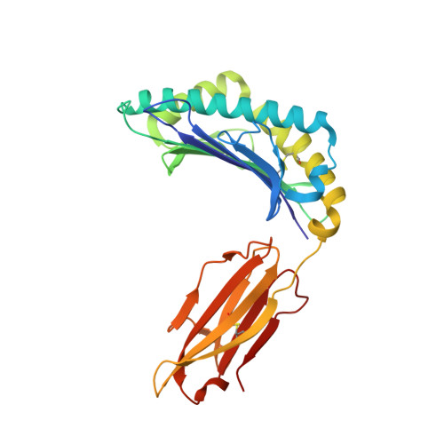
    Binding of specific antigenic peptides with human leukocyte antigen (HLA) molecules is a prerequisite for the initiation of T-cell responses and structural information about the peptide-HLA complex is essential for the detailed understanding of such interactions. HLA-A2 is the most prevalent HLA allele globally but aside from A*02:01 there is a significant lack of crystal structures, particularly for alleles that occur in high frequencies among Asian populations. Here, we report three HLA-A2 structures with the immunodominant hepatitis B core antigen 18-27 (HBcAg18-27) epitope, namely A*02:03, A*02:06, and A*02:07 at resolutions of 2.16, 1.70, and 1.75 Å respectively. This comparative analysis reveals that minor polymorphic residue changes between different HLA alleles can induce significant alterations in the major histocompatibility complex-peptide interface, and introduce conformational changes in the p3-p8 peptide region. Circular dichroism analysis demonstrated the HLA-A2-peptide complexes to have a hierarchy of thermostability and binding affinity in the order of A*02:06>A*02:07>A*02:01>A*02:03. Our findings provide structural insights into the varied HLA-A2 allele binding of the hepatitis B core antigen 18-27 epitope and the data suggest that chemical modifications of the peptide side chains could be a promising strategy to modulate and improve HLA-A2-peptide binding affinity for vaccine design.


  • Organizational Affiliation
    • Singapore Immunology Network, A*STAR, Immunos, Singapore.

Macromolecule Content 

  • Total Structure Weight: 44.83 kDa 
  • Atom Count: 3,584 
  • Modeled Residue Count: 385 
  • Deposited Residue Count: 385 
  • Unique protein chains: 3

Macromolecules

Find similar proteins by:|  3D Structure
Entity ID: 1
MoleculeChains  Sequence LengthOrganismDetailsImage
MHC class I antigen275Homo sapiensMutation(s): 0 
Gene Names: HLA-AMHC HLA-A*02:07
UniProt
Find proteins for Q861F6 (Homo sapiens)
Explore Q861F6 
Go to UniProtKB:  Q861F6
Entity Groups
Sequence Clusters30% Identity50% Identity70% Identity90% Identity95% Identity100% Identity
UniProt GroupQ861F6
Sequence Annotations
Expand
Reference Sequence
Find similar proteins by:|  3D Structure
Entity ID: 2
MoleculeChains  Sequence LengthOrganismDetailsImage
Beta-2-microglobulin100Homo sapiensMutation(s): 0 
Gene Names: B2MBETA 2-MICROGLOBULINCDABP0092HDCMA22P
UniProt & NIH Common Fund Data Resources
Find proteins for P61769 (Homo sapiens)
Explore P61769 
Go to UniProtKB:  P61769
PHAROS:  P61769
GTEx:  ENSG00000166710 
Entity Groups
Sequence Clusters30% Identity50% Identity70% Identity90% Identity95% Identity100% Identity
UniProt GroupP61769
Sequence Annotations
Expand
Reference Sequence
Find similar proteins by:  Sequence   |   3D Structure  
Entity ID: 3
MoleculeChains  Sequence LengthOrganismDetailsImage
10mer peptide from Pre-core-protein10Hepatitis B virusMutation(s): 0 
UniProt
Find proteins for P0C6H2 (Hepatitis B virus genotype C (isolate Vietnam/3270/2000))
Explore P0C6H2 
Go to UniProtKB:  P0C6H2
Entity Groups
Sequence Clusters30% Identity50% Identity70% Identity90% Identity95% Identity100% Identity
UniProt GroupP0C6H2
Sequence Annotations
Expand
Reference Sequence

Experimental Data & Validation

Experimental Data

  • Method: X-RAY DIFFRACTION
  • Resolution: 1.75 Å
  • R-Value Free:  0.207 (Depositor), 0.207 (DCC) 
  • R-Value Work:  0.177 (Depositor), 0.175 (DCC) 
  • R-Value Observed: 0.179 (Depositor) 
Space Group: P 21 21 21
Unit Cell:
Length ( Å )Angle ( ˚ )
a = 60.21α = 90
b = 79.06β = 90
c = 110γ = 90
Software Package:
Software NamePurpose
ADSCdata collection
AMoREphasing
REFMACrefinement
MOSFLMdata reduction
SCALAdata scaling

Structure Validation

View Full Validation Report


Entry History 

Deposition Data

Revision History  (Full details and data files)

  • Version 1.0: 2011-05-04
    Type: Initial release
  • Version 1.1: 2011-07-13
    Changes: Version format compliance
  • Version 1.2: 2023-11-01
    Changes: Data collection, Database references, Refinement description
  • Version 1.3: 2024-10-09
    Changes: Structure summary