3OI7 | pdb_00003oi7

Structure of the structure of the H13A mutant of Ykr043C in complex with sedoheptulose-1,7-bisphosphate


Experimental Data Snapshot

  • Method: X-RAY DIFFRACTION
  • Resolution: 2.40 Å
  • R-Value Free: 
    0.260 (Depositor), 0.260 (DCC) 
  • R-Value Work: 
    0.228 (Depositor), 0.228 (DCC) 
  • R-Value Observed: 
    0.230 (Depositor) 

Starting Model: experimental
View more details

wwPDB Validation 3D Report Full Report

Validation slider image for 3OI7

Ligand Structure Quality Assessment 


This is version 1.6 of the entry. See complete history

Literature

Riboneogenesis in yeast.

Clasquin, M.F.Melamud, E.Singer, A.Gooding, J.R.Xu, X.Dong, A.Cui, H.Campagna, S.R.Savchenko, A.Yakunin, A.F.Rabinowitz, J.D.Caudy, A.A.

(2011) Cell 145: 969-980

  • DOI: https://doi.org/10.1016/j.cell.2011.05.022
  • Primary Citation Related Structures: 
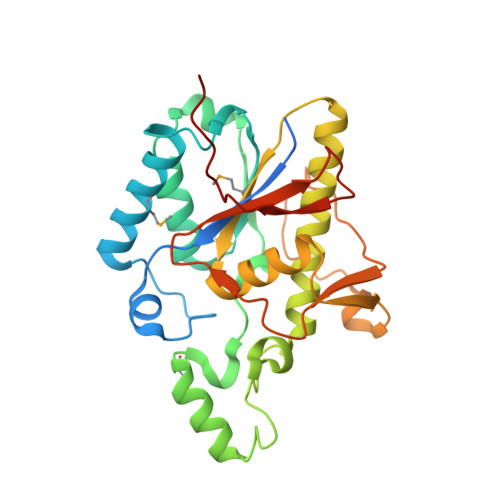
    3OI7

  • PubMed Abstract: 

    Glucose is catabolized in yeast via two fundamental routes, glycolysis and the oxidative pentose phosphate pathway, which produces NADPH and the essential nucleotide component ribose-5-phosphate. Here, we describe riboneogenesis, a thermodynamically driven pathway that converts glycolytic intermediates into ribose-5-phosphate without production of NADPH. Riboneogenesis begins with synthesis, by the combined action of transketolase and aldolase, of the seven-carbon bisphosphorylated sugar sedoheptulose-1,7-bisphosphate. In the pathway's committed step, sedoheptulose bisphosphate is hydrolyzed to sedoheptulose-7-phosphate by the enzyme sedoheptulose-1,7-bisphosphatase (SHB17), whose activity we identified based on metabolomic analysis of the corresponding knockout strain. The crystal structure of Shb17 in complex with sedoheptulose-1,7-bisphosphate reveals that the substrate binds in the closed furan form in the active site. Sedoheptulose-7-phosphate is ultimately converted by known enzymes of the nonoxidative pentose phosphate pathway to ribose-5-phosphate. Flux through SHB17 increases when ribose demand is high relative to demand for NADPH, including during ribosome biogenesis in metabolically synchronized yeast cells.


  • Organizational Affiliation
    • Lewis-Sigler Institute for Integrative Genomics, Princeton University, Princeton NJ 08544, USA.

Macromolecule Content 

  • Total Structure Weight: 136.93 kDa 
  • Atom Count: 8,833 
  • Modeled Residue Count: 1,040 
  • Deposited Residue Count: 1,168 
  • Unique protein chains: 1

Macromolecules

Find similar proteins by:|  3D Structure
Entity ID: 1
MoleculeChains  Sequence LengthOrganismDetailsImage
Uncharacterized protein YKR043C
A, B, C, D
292Saccharomyces cerevisiaeMutation(s): 1 
Gene Names: YKR043C
EC: 3.1.3.11 (PDB Primary Data), 3.1.3.37 (UniProt)
UniProt
Find proteins for P36136 (Saccharomyces cerevisiae (strain ATCC 204508 / S288c))
Explore P36136 
Go to UniProtKB:  P36136
Entity Groups
Sequence Clusters30% Identity50% Identity70% Identity90% Identity95% Identity100% Identity
UniProt GroupP36136
Sequence Annotations
Expand
Reference Sequence

Small Molecules

Ligands 5 Unique
IDChains Name / Formula / InChI Key2D Diagram3D Interactions
OI7

Query on OI7


Download:Ideal Coordinates CCD File
E [auth A],
K [auth B],
O [auth C],
U [auth D]
1,7-di-O-phosphono-beta-D-altro-hept-2-ulofuranose
C7 H16 O13 P2
YCQNLCZEHKXXNQ-BNWJMWRWSA-N
GOL

Query on GOL


Download:Ideal Coordinates CCD File
J [auth A]GLYCEROL
C3 H8 O3
PEDCQBHIVMGVHV-UHFFFAOYSA-N
EDO

Query on EDO


Download:Ideal Coordinates CCD File
H [auth A]
I [auth A]
N [auth B]
S [auth C]
T [auth C]
H [auth A],
I [auth A],
N [auth B],
S [auth C],
T [auth C],
X [auth D],
Y [auth D],
Z [auth D]
1,2-ETHANEDIOL
C2 H6 O2
LYCAIKOWRPUZTN-UHFFFAOYSA-N
MG

Query on MG


Download:Ideal Coordinates CCD File
F [auth A],
L [auth B],
P [auth C],
V [auth D]
MAGNESIUM ION
Mg
JLVVSXFLKOJNIY-UHFFFAOYSA-N
NA

Query on NA


Download:Ideal Coordinates CCD File
G [auth A],
M [auth B],
Q [auth C],
R [auth C],
W [auth D]
SODIUM ION
Na
FKNQFGJONOIPTF-UHFFFAOYSA-N
Modified Residues  1 Unique
IDChains TypeFormula2D DiagramParent
MSE
Query on MSE
A, B, C, D
L-PEPTIDE LINKINGC5 H11 N O2 SeMET

Experimental Data & Validation

Experimental Data

  • Method: X-RAY DIFFRACTION
  • Resolution: 2.40 Å
  • R-Value Free:  0.260 (Depositor), 0.260 (DCC) 
  • R-Value Work:  0.228 (Depositor), 0.228 (DCC) 
  • R-Value Observed: 0.230 (Depositor) 
Space Group: P 1
Unit Cell:
Length ( Å )Angle ( ˚ )
a = 58.559α = 90.04
b = 74.945β = 89.92
c = 83.615γ = 77.22
Software Package:
Software NamePurpose
CrystalCleardata collection
PHASERphasing
REFMACrefinement
HKL-2000data reduction
HKL-2000data scaling

Structure Validation

View Full Validation Report


Ligand Structure Quality Assessment 


Entry History 

Revision History  (Full details and data files)

  • Version 1.0: 2010-11-17
    Type: Initial release
  • Version 1.1: 2011-07-13
    Changes: Version format compliance
  • Version 1.2: 2011-08-24
    Changes: Database references
  • Version 1.3: 2020-07-29
    Type: Remediation
    Reason: Carbohydrate remediation
    Changes: Data collection, Database references, Derived calculations
  • Version 1.4: 2023-09-06
    Changes: Data collection, Database references, Refinement description, Structure summary
  • Version 1.5: 2023-12-06
    Changes: Data collection
  • Version 1.6: 2024-11-20
    Changes: Structure summary