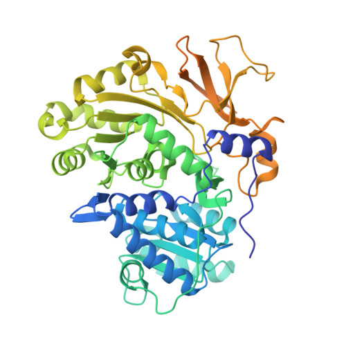
3O83

Structure of BasE N-terminal domain from Acinetobacter baumannii bound to 2-(4-n-dodecyl-1,2,3-triazol-1-yl)-5'-O-[N-(2-hydroxybenzoyl)sulfamoyl]adenosine


Experimental Data Snapshot

  • Method: X-RAY DIFFRACTION
  • Resolution: 1.90 Å
  • R-Value Free: 0.217 
  • R-Value Work: 0.194 
  • R-Value Observed: 0.195 

wwPDB Validation   3D Report Full Report


This is version 1.2 of the entry. See complete history


Literature

Biochemical and structural characterization of bisubstrate inhibitors of BasE, the self-standing nonribosomal peptide synthetase adenylate-forming enzyme of acinetobactin synthesis.

Drake, E.J.Duckworth, B.P.Neres, J.Aldrich, C.C.Gulick, A.M.

(2010) Biochemistry 49: 9292-9305

  • DOI: https://doi.org/10.1021/bi101226n
  • Primary Citation of Related Structures:  
    3O82, 3O83, 3O84

  • PubMed Abstract: 

    The human pathogen Acinetobacter baumannii produces a siderophore called acinetobactin that is derived from one molecule each of threonine, histidine, and 2,3-dihydroxybenzoic acid (DHB). The activity of several nonribosomal peptide synthetase (NRPS) enzymes is used to combine the building blocks into the final molecule. The acinetobactin synthesis pathway initiates with a self-standing adenylation enzyme, BasE, that activates the DHB molecule and covalently transfers it to the pantetheine cofactor of an aryl-carrier protein of BasF, a strategy that is shared with many siderophore-producing NRPS clusters. In this reaction, DHB reacts with ATP to form the aryl adenylate and pyrophosphate. In a second partial reaction, the DHB is transferred to the carrier protein. Inhibitors of BasE and related enzymes have been identified that prevent growth of bacteria on iron-limiting media. Recently, a new inhibitor of BasE has been identified via high-throughput screening using a fluorescence polarization displacement assay. We present here biochemical and structural studies to examine the binding mode of this inhibitor. The kinetics of the wild-type BasE enzyme is shown, and inhibition studies demonstrate that the new compound exhibits competitive inhibition against both ATP and 2,3-dihydroxybenzoate. Structural examination of BasE bound to this inhibitor illustrates a novel binding mode in which the phenyl moiety partially fills the enzyme pantetheine binding tunnel. Structures of rationally designed bisubstrate inhibitors are also presented.


  • Organizational Affiliation

    Hauptman-Woodward Institute and Department of Structural Biology, University at Buffalo, Buffalo, NY 14203-1102, USA.


Macromolecules
Find similar proteins by:  (by identity cutoff)  |  3D Structure
Entity ID: 1
MoleculeChains Sequence LengthOrganismDetailsImage
Peptide arylation enzyme
A, B
544Acinetobacter baumannii AB900Mutation(s): 1 
Gene Names: ACICU_02578basE
EC: 6.2.1
Entity Groups  
Sequence Clusters30% Identity50% Identity70% Identity90% Identity95% Identity100% Identity
Sequence Annotations
Expand
  • Reference Sequence
Small Molecules
Ligands 4 Unique
IDChains Name / Formula / InChI Key2D Diagram3D Interactions
IXN
Query on IXN

Download Ideal Coordinates CCD File 
C [auth A],
H [auth B]
2-(4-dodecyl-1H-1,2,3-triazol-1-yl)-5'-O-{[(2-hydroxyphenyl)carbonyl]sulfamoyl}adenosine
C31 H43 N9 O8 S
IZIYHQKZTVZLLO-NYBSAPDNSA-N
MRD
Query on MRD

Download Ideal Coordinates CCD File 
L [auth B](4R)-2-METHYLPENTANE-2,4-DIOL
C6 H14 O2
SVTBMSDMJJWYQN-RXMQYKEDSA-N
MPD
Query on MPD

Download Ideal Coordinates CCD File 
G [auth A](4S)-2-METHYL-2,4-PENTANEDIOL
C6 H14 O2
SVTBMSDMJJWYQN-YFKPBYRVSA-N
CA
Query on CA

Download Ideal Coordinates CCD File 
D [auth A]
E [auth A]
F [auth A]
I [auth B]
J [auth B]
D [auth A],
E [auth A],
F [auth A],
I [auth B],
J [auth B],
K [auth B]
CALCIUM ION
Ca
BHPQYMZQTOCNFJ-UHFFFAOYSA-N
Experimental Data & Validation

Experimental Data

  • Method: X-RAY DIFFRACTION
  • Resolution: 1.90 Å
  • R-Value Free: 0.217 
  • R-Value Work: 0.194 
  • R-Value Observed: 0.195 
  • Space Group: P 21 21 21
Unit Cell:
Length ( Å )Angle ( ˚ )
a = 66.46α = 90
b = 144.238β = 90
c = 148.469γ = 90
Software Package:
Software NamePurpose
Blu-Icedata collection
MOLREPphasing
REFMACrefinement
HKL-2000data reduction
HKL-2000data scaling

Structure Validation

View Full Validation Report


Entry History 

Deposition Data

Revision History  (Full details and data files)

  • Version 1.0: 2010-10-06
    Type: Initial release
  • Version 1.1: 2011-07-13
    Changes: Version format compliance
  • Version 1.2: 2023-09-06
    Changes: Data collection, Database references, Derived calculations, Refinement description