3O79 | pdb_00003o79

Crystal Structure of Wild-type Rabbit PrP 126-230


Experimental Data Snapshot

  • Method: X-RAY DIFFRACTION
  • Resolution: 1.60 Å
  • R-Value Free: 
    0.218 (Depositor), 0.238 (DCC) 
  • R-Value Work: 
    0.157 (Depositor) 
  • R-Value Observed: 
    0.161 (Depositor) 

Starting Model: experimental
View more details

wwPDB Validation 3D Report Full Report

Validation slider image for 3O79

This is version 1.4 of the entry. See complete history

Literature

Prion disease susceptibility is affected by beta-structure folding propensity and local side-chain interactions in PrP.

Khan, M.Q.Sweeting, B.Mulligan, V.K.Arslan, P.E.Cashman, N.R.Pai, E.F.Chakrabartty, A.

(2010) Proc Natl Acad Sci U S A 107: 19808-19813

  • DOI: https://doi.org/10.1073/pnas.1005267107
  • Primary Citation Related Structures: 
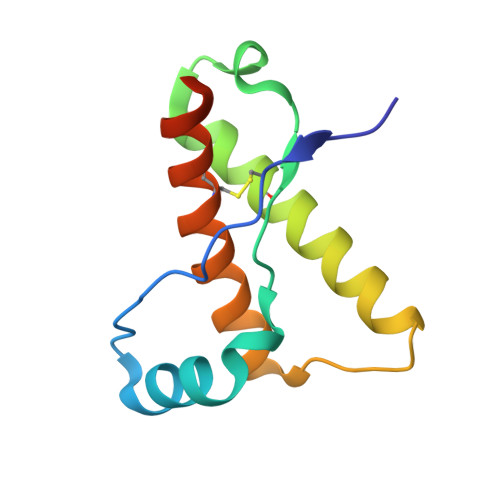
    3O79

  • PubMed Abstract: 

    Prion diseases occur when the normally α-helical prion protein (PrP) converts to a pathological β-structured state with prion infectivity (PrP(Sc)). Exposure to PrP(Sc) from other mammals can catalyze this conversion. Evidence from experimental and accidental transmission of prions suggests that mammals vary in their prion disease susceptibility: Hamsters and mice show relatively high susceptibility, whereas rabbits, horses, and dogs show low susceptibility. Using a novel approach to quantify conformational states of PrP by circular dichroism (CD), we find that prion susceptibility tracks with the intrinsic propensity of mammalian PrP to convert from the native, α-helical state to a cytotoxic β-structured state, which exists in a monomer-octamer equilibrium. It has been controversial whether β-structured monomers exist at acidic pH; sedimentation equilibrium and dual-wavelength CD evidence is presented for an equilibrium between a β-structured monomer and octamer in some acidic pH conditions. Our X-ray crystallographic structure of rabbit PrP has identified a key helix-capping motif implicated in the low prion disease susceptibility of rabbits. Removal of this capping motif increases the β-structure folding propensity of rabbit PrP to match that of PrP from mouse, a species more susceptible to prion disease.


  • Organizational Affiliation
    • Campbell Family Institute for Cancer Research, Department of Biochemistry, University of Toronto, Toronto Medical Discovery Tower 4-307, 101 College Street, Toronto, ON, Canada M5G 1L7.

Macromolecule Content 

  • Total Structure Weight: 25.32 kDa 
  • Atom Count: 1,960 
  • Modeled Residue Count: 202 
  • Deposited Residue Count: 210 
  • Unique protein chains: 1

Macromolecules

Find similar proteins by:|  3D Structure
Entity ID: 1
MoleculeChains  Sequence LengthOrganismDetailsImage
Rabbit PrP
A, B
105Oryctolagus cuniculusMutation(s): 0 
Gene Names: PRNP
UniProt
Find proteins for Q9TSF8 (Oryctolagus cuniculus)
Explore Q9TSF8 
Go to UniProtKB:  Q9TSF8
Entity Groups
Sequence Clusters30% Identity50% Identity70% Identity90% Identity95% Identity100% Identity
UniProt GroupQ9TSF8
Sequence Annotations
Expand
Reference Sequence

Small Molecules

Ligands 3 Unique
IDChains Name / Formula / InChI Key2D Diagram3D Interactions
GOL

Query on GOL


Download:Ideal Coordinates CCD File
I [auth B],
J [auth B],
K [auth B]
GLYCEROL
C3 H8 O3
PEDCQBHIVMGVHV-UHFFFAOYSA-N
CL

Query on CL


Download:Ideal Coordinates CCD File
E [auth A],
H [auth B]
CHLORIDE ION
Cl
VEXZGXHMUGYJMC-UHFFFAOYSA-M
NA

Query on NA


Download:Ideal Coordinates CCD File
C [auth A],
D [auth A],
F [auth B],
G [auth B]
SODIUM ION
Na
FKNQFGJONOIPTF-UHFFFAOYSA-N

Experimental Data & Validation

Experimental Data

  • Method: X-RAY DIFFRACTION
  • Resolution: 1.60 Å
  • R-Value Free:  0.218 (Depositor), 0.238 (DCC) 
  • R-Value Work:  0.157 (Depositor) 
  • R-Value Observed: 0.161 (Depositor) 
Space Group: P 21 21 21
Unit Cell:
Length ( Å )Angle ( ˚ )
a = 29.53α = 90
b = 86.17β = 90
c = 87.14γ = 90
Software Package:
Software NamePurpose
XSCALEdata scaling
REFMACrefinement
PDB_EXTRACTdata extraction
Macromoleculardata collection
XDSdata reduction
PHASERphasing

Structure Validation

View Full Validation Report


Entry History 

Deposition Data

Revision History  (Full details and data files)

  • Version 1.0: 2010-11-24
    Type: Initial release
  • Version 1.1: 2011-07-13
    Changes: Version format compliance
  • Version 1.2: 2023-09-06
    Changes: Data collection, Database references, Derived calculations, Refinement description
  • Version 1.3: 2024-03-13
    Changes: Source and taxonomy
  • Version 1.4: 2024-10-16
    Changes: Structure summary