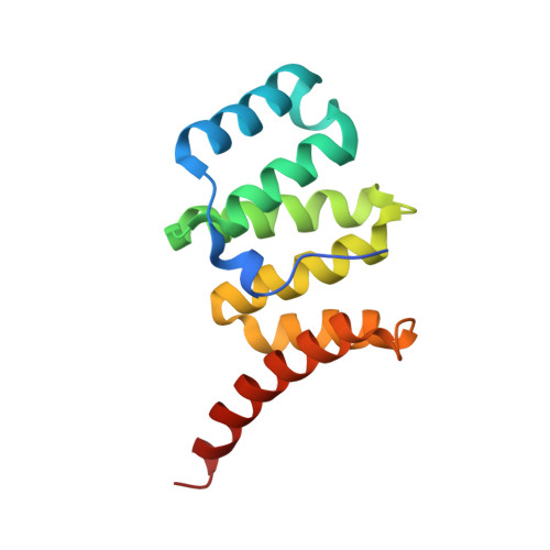
3O48

Crystal structure of fission protein Fis1 from Saccharomyces cerevisiae


Experimental Data Snapshot

  • Method: X-RAY DIFFRACTION
  • Resolution: 1.75 Å
  • R-Value Free: 0.188 
  • R-Value Work: 0.165 
  • R-Value Observed: 0.166 

wwPDB Validation   3D Report Full Report


This is version 1.2 of the entry. See complete history


Literature

The 1.75 Angstrom resolution structure of fission protein Fis1 from Saccharomyces cerevisiae reveals elusive interactions of the autoinhibitory domain

Tooley, J.E.Khangulov, V.Lees, J.P.Schlessman, J.L.Bewley, M.C.Heroux, A.Bosch, J.Hill, R.B.

(2011) Acta Crystallogr Sect F Struct Biol Cryst Commun 67: 1310-1315

  • DOI: https://doi.org/10.1107/S1744309111029368
  • Primary Citation of Related Structures:  
    3O48

  • PubMed Abstract: 

    Fis1 mediates mitochondrial and peroxisomal fission. It is tail-anchored to these organelles by a transmembrane domain, exposing a soluble cytoplasmic domain. Previous studies suggested that Fis1 is autoinhibited by its N-terminal region. Here, a 1.75 Å resolution crystal structure of the Fis1 cytoplasmic domain from Saccharomyces cerevisiae is reported which adopts a tetratricopeptide-repeat fold. It is observed that this fold creates a concave surface important for fission, but is sterically occluded by its N-terminal region. Thus, this structure provides a physical basis for autoinhibition and allows a detailed examination of the interactions that stabilize the inhibited state of this molecule.


  • Organizational Affiliation

    Department of Biology, Johns Hopkins University, Baltimore, MD 21218, USA.


Macromolecules
Find similar proteins by:  (by identity cutoff)  |  3D Structure
Entity ID: 1
MoleculeChains Sequence LengthOrganismDetailsImage
Mitochondria fission 1 protein134Saccharomyces cerevisiaeMutation(s): 0 
Gene Names: FIS1MDV2YIL065C
UniProt
Find proteins for P40515 (Saccharomyces cerevisiae (strain ATCC 204508 / S288c))
Explore P40515 
Go to UniProtKB:  P40515
Entity Groups  
Sequence Clusters30% Identity50% Identity70% Identity90% Identity95% Identity100% Identity
UniProt GroupP40515
Sequence Annotations
Expand
  • Reference Sequence
Experimental Data & Validation

Experimental Data

  • Method: X-RAY DIFFRACTION
  • Resolution: 1.75 Å
  • R-Value Free: 0.188 
  • R-Value Work: 0.165 
  • R-Value Observed: 0.166 
  • Space Group: P 43 21 2
Unit Cell:
Length ( Å )Angle ( ˚ )
a = 46.02α = 90
b = 46.02β = 90
c = 139.22γ = 90
Software Package:
Software NamePurpose
HKL-2000data collection
BALBESphasing
REFMACrefinement
XDSdata reduction
XSCALEdata scaling

Structure Validation

View Full Validation Report


Entry History 

Deposition Data

Revision History  (Full details and data files)

  • Version 1.0: 2011-08-10
    Type: Initial release
  • Version 1.1: 2011-12-28
    Changes: Database references
  • Version 1.2: 2023-09-06
    Changes: Data collection, Database references, Refinement description