3O0D | pdb_00003o0d

Crystal structure of Lip2 lipase from Yarrowia lipolytica at 1.7 A resolution


Experimental Data Snapshot

  • Method: X-RAY DIFFRACTION
  • Resolution: 1.70 Å
  • R-Value Free: 
    0.211 (Depositor), 0.210 (DCC) 
  • R-Value Work: 
    0.171 (Depositor), 0.170 (DCC) 
  • R-Value Observed: 
    0.173 (Depositor) 

wwPDB Validation 3D Report Full Report

Validation slider image for 3O0D

Ligand Structure Quality Assessment 


This is version 1.3 of the entry. See complete history

Literature

Exploring the conformational states and rearrangements of Yarrowia lipolytica Lipase.

Bordes, F.Barbe, S.Escalier, P.Mourey, L.Andre, I.Marty, A.Tranier, S.

(2010) Biophys J 99: 2225-2234

  • DOI: https://doi.org/10.1016/j.bpj.2010.07.040
  • Primary Citation Related Structures: 
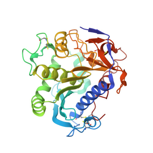
    3O0D

  • PubMed Abstract: 

    We report the 1.7 Å resolution crystal structure of the Lip2 lipase from Yarrowia lipolytica in its closed conformation. The Lip2 structure is highly homologous to known structures of the fungal lipase family (Thermomyces lanuginosa, Rhizopus niveus, and Rhizomucor miehei lipases). However, it also presents some unique features that are described and discussed here in detail. Structural differences, in particular in the conformation adopted by the so-called lid subdomain, suggest that the opening mechanism of Lip2 may differ from that of other fungal lipases. Because the catalytic activity of lipases is strongly dependent on structural rearrangement of this mobile subdomain, we focused on elucidating the molecular mechanism of lid motion. Using the x-ray structure of Lip2, we carried out extensive molecular-dynamics simulations in explicit solvent environments (water and water/octane interface) to characterize the major structural rearrangements that the lid undergoes under the influence of solvent or upon substrate binding. Overall, our results suggest a two-step opening mechanism that gives rise first to a semi-open conformation upon adsorption of the protein at the water/organic solvent interface, followed by a further opening of the lid upon substrate binding.


  • Organizational Affiliation
    • Université de Toulouse, Toulouse, France.

Macromolecule Content 

  • Total Structure Weight: 239.29 kDa 
  • Atom Count: 18,591 
  • Modeled Residue Count: 2,095 
  • Deposited Residue Count: 2,107 
  • Unique protein chains: 1

Macromolecules

Find similar proteins by:|  3D Structure
Entity ID: 1
MoleculeChains  Sequence LengthOrganismDetailsImage
Triacylglycerol lipase
A, B, C, D, E
A, B, C, D, E, F, G
301Yarrowia lipolyticaMutation(s): 0 
Gene Names: lip2YALI0A20350g
EC: 3.1.1.3
Membrane Entity: Yes 
UniProt
Find proteins for Q9P8F7 (Yarrowia lipolytica)
Explore Q9P8F7 
Go to UniProtKB:  Q9P8F7
Entity Groups
Sequence Clusters30% Identity50% Identity70% Identity90% Identity95% Identity100% Identity
UniProt GroupQ9P8F7
Glycosylation
Glycosylation Sites: 2
Sequence Annotations
Expand
Reference Sequence

Small Molecules

Ligands 4 Unique
IDChains Name / Formula / InChI Key2D Diagram3D Interactions
NAG

Query on NAG


Download:Ideal Coordinates CCD File
AA [auth D]
BA [auth D]
GA [auth E]
H [auth A]
HA [auth E]
AA [auth D],
BA [auth D],
GA [auth E],
H [auth A],
HA [auth E],
I [auth A],
LA [auth F],
MA [auth F],
O [auth B],
P [auth B],
QA [auth G],
RA [auth G],
U [auth C],
V [auth C]
2-acetamido-2-deoxy-beta-D-glucopyranose
C8 H15 N O6
OVRNDRQMDRJTHS-FMDGEEDCSA-N
MPD

Query on MPD


Download:Ideal Coordinates CCD File
CA [auth D]
EA [auth D]
IA [auth E]
J [auth A]
JA [auth E]
CA [auth D],
EA [auth D],
IA [auth E],
J [auth A],
JA [auth E],
K [auth A],
L [auth A],
OA [auth F],
Q [auth B],
R [auth B],
W [auth C],
X [auth C],
Y [auth C]
(4S)-2-METHYL-2,4-PENTANEDIOL
C6 H14 O2
SVTBMSDMJJWYQN-YFKPBYRVSA-N
MRD

Query on MRD


Download:Ideal Coordinates CCD File
DA [auth D],
M [auth A],
NA [auth F],
S [auth B]
(4R)-2-METHYLPENTANE-2,4-DIOL
C6 H14 O2
SVTBMSDMJJWYQN-RXMQYKEDSA-N
K

Query on K


Download:Ideal Coordinates CCD File
FA [auth D]
KA [auth E]
N [auth A]
PA [auth F]
SA [auth G]
FA [auth D],
KA [auth E],
N [auth A],
PA [auth F],
SA [auth G],
T [auth B],
Z [auth C]
POTASSIUM ION
K
NPYPAHLBTDXSSS-UHFFFAOYSA-N

Experimental Data & Validation

Experimental Data

  • Method: X-RAY DIFFRACTION
  • Resolution: 1.70 Å
  • R-Value Free:  0.211 (Depositor), 0.210 (DCC) 
  • R-Value Work:  0.171 (Depositor), 0.170 (DCC) 
  • R-Value Observed: 0.173 (Depositor) 
Space Group: P 21 21 21
Unit Cell:
Length ( Å )Angle ( ˚ )
a = 115.354α = 90
b = 132.141β = 90
c = 137.255γ = 90
Software Package:
Software NamePurpose
DNAdata collection
MOLREPphasing
REFMACrefinement
MOSFLMdata reduction
SCALAdata scaling

Structure Validation

View Full Validation Report


Ligand Structure Quality Assessment 


Entry History 

Deposition Data

Revision History  (Full details and data files)

  • Version 1.0: 2010-11-24
    Type: Initial release
  • Version 1.1: 2011-07-13
    Changes: Version format compliance
  • Version 1.2: 2020-07-29
    Type: Remediation
    Reason: Carbohydrate remediation
    Changes: Data collection, Derived calculations, Structure summary
  • Version 1.3: 2024-11-20
    Changes: Data collection, Database references, Structure summary