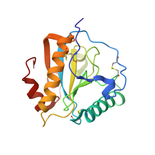
3NW3

Crystal structure of the complex of peptidoglycan recognition protein (PGRP-S) with the PGN Fragment at 2.5 A resolution


Experimental Data Snapshot

  • Method: X-RAY DIFFRACTION
  • Resolution: 2.50 Å
  • R-Value Free: 0.249 
  • R-Value Work: 0.236 
  • R-Value Observed: 0.236 

wwPDB Validation   3D Report Full Report


Ligand Structure Quality Assessment 


This is version 4.0 of the entry. See complete history


Literature

Multiligand specificity of pathogen-associated molecular pattern-binding site in peptidoglycan recognition protein

Sharma, P.Dube, D.Sinha, M.Mishra, B.Dey, S.Mal, G.Pathak, K.M.Kaur, P.Sharma, S.Singh, T.P.

(2011) J Biol Chem 286: 31723-31730

  • DOI: https://doi.org/10.1074/jbc.M111.264374
  • Primary Citation of Related Structures:  
    3NG4, 3NW3

  • PubMed Abstract: 

    The peptidoglycan recognition protein PGRP-S is an innate immunity molecule that specifically interacts with microbial peptidoglycans and other pathogen-associated molecular patterns. We report here two structures of the unique tetrameric camel PGRP-S (CPGRP-S) complexed with (i) muramyl dipeptide (MDP) at 2.5 Å resolution and (ii) GlcNAc and β-maltose at 1.7Å resolution. The binding studies carried out using surface plasmon resonance indicated that CPGRP-S binds to MDP with a dissociation constant of 10(-7) M, whereas the binding affinities for GlcNAc and β-maltose separately are in the range of 10(-4) M to 10(-5) M, whereas the dissociation constant for the mixture of GlcNAc and maltose was estimated to be 10(-6) M. The data from bacterial suspension culture experiments showed a significant inhibition of the growth of Staphylococcus aureus cells when CPGRP-S was added to culture medium. The ELISA experiment showed that the amount of MDP-induced production of TNF-α and IL-6 decreased considerably after the introduction of CPGRP-S. The crystal structure determinations of (i) a binary complex with MDP and (ii) a ternary complex with GlcNAc and β-maltose revealed that MDP, GlcNAc, and β-maltose bound to CPGRP-S in the ligand binding cleft, which is situated at the interface of molecules C and D of the homotetramer formed by four protein molecules A, B, C, and D. In the binary complex, the muramyl moiety of MDP is observed at the C-D interface, whereas the peptide chain protrudes into the center of tetramer. In the ternary complex, GlcNAc and β-maltose occupy distinct non-overlapping positions belonging to different subsites.


  • Organizational Affiliation

    Department of Biophysics, All India Institute of Medical Sciences, New Delhi, India 110029.


Macromolecules
Find similar proteins by:  (by identity cutoff)  |  3D Structure
Entity ID: 1
MoleculeChains Sequence LengthOrganismDetailsImage
Peptidoglycan recognition protein 1
A, B, C, D
171Camelus dromedariusMutation(s): 0 
UniProt
Find proteins for Q9GK12 (Camelus dromedarius)
Explore Q9GK12 
Go to UniProtKB:  Q9GK12
Entity Groups  
Sequence Clusters30% Identity50% Identity70% Identity90% Identity95% Identity100% Identity
UniProt GroupQ9GK12
Sequence Annotations
Expand
  • Reference Sequence
Small Molecules
Ligands 6 Unique
IDChains Name / Formula / InChI Key2D Diagram3D Interactions
NDG
Query on NDG

Download Ideal Coordinates CCD File 
E [auth C]2-acetamido-2-deoxy-alpha-D-glucopyranose
C8 H15 N O6
OVRNDRQMDRJTHS-PVFLNQBWSA-N
TLA
Query on TLA

Download Ideal Coordinates CCD File 
F [auth D]L(+)-TARTARIC ACID
C4 H6 O6
FEWJPZIEWOKRBE-JCYAYHJZSA-N
DGN
Query on DGN

Download Ideal Coordinates CCD File 
H [auth D]D-GLUTAMINE
C5 H10 N2 O3
ZDXPYRJPNDTMRX-GSVOUGTGSA-N
GOL
Query on GOL

Download Ideal Coordinates CCD File 
G [auth D]GLYCEROL
C3 H8 O3
PEDCQBHIVMGVHV-UHFFFAOYSA-N
LAC
Query on LAC

Download Ideal Coordinates CCD File 
J [auth D]LACTIC ACID
C3 H6 O3
JVTAAEKCZFNVCJ-UWTATZPHSA-N
ALA
Query on ALA

Download Ideal Coordinates CCD File 
I [auth D]ALANINE
C3 H7 N O2
QNAYBMKLOCPYGJ-REOHCLBHSA-N
Experimental Data & Validation

Experimental Data

  • Method: X-RAY DIFFRACTION
  • Resolution: 2.50 Å
  • R-Value Free: 0.249 
  • R-Value Work: 0.236 
  • R-Value Observed: 0.236 
  • Space Group: I 2 2 2
Unit Cell:
Length ( Å )Angle ( ˚ )
a = 87.17α = 90
b = 102β = 90
c = 161.6γ = 90
Software Package:
Software NamePurpose
DENZOdata reduction
AMoREphasing
CNSrefinement
AUTOMARdata reduction
SCALEPACKdata scaling

Structure Validation

View Full Validation Report


Ligand Structure Quality Assessment 


Entry History 

Deposition Data

Revision History  (Full details and data files)

  • Version 1.0: 2010-08-04
    Type: Initial release
  • Version 1.1: 2011-07-13
    Changes: Version format compliance
  • Version 1.2: 2012-06-13
    Changes: Database references
  • Version 2.0: 2020-07-01
    Changes: Advisory, Atomic model, Data collection, Derived calculations, Non-polymer description, Structure summary
  • Version 3.0: 2020-07-29
    Type: Remediation
    Reason: Carbohydrate remediation
    Changes: Atomic model, Data collection, Derived calculations, Structure summary
  • Version 3.1: 2023-11-01
    Changes: Data collection, Database references, Refinement description, Structure summary
  • Version 4.0: 2023-11-15
    Changes: Atomic model, Data collection, Derived calculations