3NTO

Crystal structure of K97V mutant myo-inositol dehydrogenase from Bacillus subtilis


Experimental Data Snapshot

  • Method: X-RAY DIFFRACTION
  • Resolution: 1.91 Å
  • R-Value Free: 0.209 
  • R-Value Work: 0.168 
  • R-Value Observed: 0.170 

wwPDB Validation   3D Report Full Report


This is version 1.4 of the entry. See complete history


Literature

Structural investigation of myo-inositol dehydrogenase from Bacillus subtilis: implications for catalytic mechanism and inositol dehydrogenase subfamily classification.

van Straaten, K.E.Zheng, H.Palmer, D.R.Sanders, D.A.

(2010) Biochem J 432: 237-247

  • DOI: https://doi.org/10.1042/BJ20101079
  • Primary Citation of Related Structures:  
    3MZ0, 3NT2, 3NT4, 3NT5, 3NTO, 3NTQ, 3NTR

  • PubMed Abstract: 

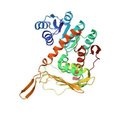
    Inositol dehydrogenase from Bacillus subtilis (BsIDH) is a NAD+-dependent enzyme that catalyses the oxidation of the axial hydroxy group of myo-inositol to form scyllo-inosose. We have determined the crystal structures of wild-type BsIDH and of the inactive K97V mutant in apo-, holo- and ternary complexes with inositol and inosose. BsIDH is a tetramer, with a novel arrangement consisting of two long continuous β-sheets, formed from all four monomers, in which the two central strands are crossed over to form the core of the tetramer. Each subunit in the tetramer consists of two domains: an N-terminal Rossmann fold domain containing the cofactor-binding site, and a C-terminal domain containing the inositol-binding site. Structural analysis allowed us to determine residues important in cofactor and substrate binding. Lys97, Asp172 and His176 are the catalytic triad involved in the catalytic mechanism of BsIDH, similar to what has been proposed for related enzymes and short-chain dehydrogenases. Furthermore, a conformational change in the nicotinamide ring was observed in some ternary complexes, suggesting hydride transfer to the si-face of NAD+. Finally, comparison of the structure and sequence of BsIDH with other putative inositol dehydrogenases allowed us to differentiate these enzymes into four subfamilies based on six consensus sequence motifs defining the cofactor- and substrate-binding sites.


  • Organizational Affiliation

    Department of Chemistry, University of Saskatchewan, Saskatoon, SK, Canada, S7N 5C9.


Macromolecules
Find similar proteins by:  (by identity cutoff)  |  3D Structure
Entity ID: 1
MoleculeChains Sequence LengthOrganismDetailsImage
Inositol 2-dehydrogenase/D-chiro-inositol 3-dehydrogenase344Bacillus subtilisMutation(s): 1 
Gene Names: BSU39700E83GidhiolGNP_391849.2
EC: 1.1.1.18
UniProt
Find proteins for P26935 (Bacillus subtilis (strain 168))
Explore P26935 
Go to UniProtKB:  P26935
Entity Groups  
Sequence Clusters30% Identity50% Identity70% Identity90% Identity95% Identity100% Identity
UniProt GroupP26935
Sequence Annotations
Expand
  • Reference Sequence
Small Molecules
Ligands 1 Unique
IDChains Name / Formula / InChI Key2D Diagram3D Interactions
EPE
Query on EPE

Download Ideal Coordinates CCD File 
B [auth A]4-(2-HYDROXYETHYL)-1-PIPERAZINE ETHANESULFONIC ACID
C8 H18 N2 O4 S
JKMHFZQWWAIEOD-UHFFFAOYSA-N
Experimental Data & Validation

Experimental Data

  • Method: X-RAY DIFFRACTION
  • Resolution: 1.91 Å
  • R-Value Free: 0.209 
  • R-Value Work: 0.168 
  • R-Value Observed: 0.170 
  • Space Group: I 2 2 2
Unit Cell:
Length ( Å )Angle ( ˚ )
a = 52.27α = 90
b = 119.5β = 90
c = 128.7γ = 90
Software Package:
Software NamePurpose
XSCALEdata scaling
MOLREPphasing
PHENIXrefinement
PDB_EXTRACTdata extraction
MxDCdata collection
XDSdata reduction

Structure Validation

View Full Validation Report


Entry History 

Deposition Data

Revision History  (Full details and data files)

  • Version 1.0: 2010-09-15
    Type: Initial release
  • Version 1.1: 2011-07-13
    Changes: Version format compliance
  • Version 1.2: 2017-11-08
    Changes: Refinement description
  • Version 1.3: 2023-12-27
    Changes: Data collection, Database references, Derived calculations
  • Version 1.4: 2024-04-03
    Changes: Refinement description