Halogen-substituted (C-beta-D-glucopyranosyl)-hydroquinone regioisomers: synthesis, enzymatic evaluation and their binding to glycogen phosphorylase.
Alexacou, K.M., Zhang, Y.Z., Praly, J.P., Zographos, S.E., Chrysina, E.D., Oikonomakos, N.G., Leonidas, D.D.(2011) Bioorg Med Chem 19: 5125-5136
- PubMed: 21821421 Search on PubMed
- DOI: https://doi.org/10.1016/j.bmc.2011.07.024
- Primary Citation Related Structures:
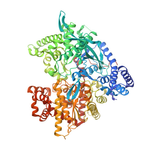
3NP7, 3NP9, 3NPA, 3S0J - PubMed Abstract:
Electrophilic halogenation of C-(2,3,4,6-tetra-O-acetyl-β-D-glucopyranosyl) 1,4-dimethoxybenzene (1) afforded regioselectively products halogenated at the para position to the D-glucosyl moiety (8, 9) that were deacetylated to 3 (chloride) and 16 (bromide). For preparing meta regioisomers, 1 was efficiently oxidized with CAN to afford C-(2,3,4,6-tetra-O-acetyl-β-D-glucopyranosyl) 1,4-benzoquinone 2 which, in either MeOH or H(2)O-THF containing few equivalents of AcCl, added hydrochloric acid to produce predominantly meta (with respect to the sugar moiety) chlorinated hydroquinone derivatives 5 and 18, this latter being deacetylated to 4. The deacetylated meta (4, 5) or para (3, 16) halohydroquinones were evaluated as inhibitors of glycogen phosphorylase (GP, a molecular target for inhibition of hepatic glycogenolysis under high glucose concentrations) by kinetics and X-ray crystallography. These compounds are competitive inhibitors of GPb with respect to α-D-glucose-1-phosphate. The measured IC(50) values (μM) [169.9±10.0 (3), 95 (4), 39.8±0.3 (5) 136.4±4.9 (16)] showed that the meta halogenated inhibitors (4, 5) are more potent than their para analogs (3, 16). The crystal structures of GPb in complex with these compounds at high resolution (1.97-2.05 Å) revealed that the inhibitors are accommodated at the catalytic site and stabilize the T conformation of the enzyme. The differences in their inhibitory potency can be interpreted in terms of variations in the interactions with protein residues of the different substituents on the aromatic part of the inhibitors.
- Institute of Organic and Pharmaceutical Chemistry, National Hellenic Research Foundation, Athens, Greece.
Organizational Affiliation: