3N89 | pdb_00003n89

KH domains


Experimental Data Snapshot

  • Method: X-RAY DIFFRACTION
  • Resolution: 2.79 Å
  • R-Value Free: 
    0.276 (Depositor), 0.275 (DCC) 
  • R-Value Work: 
    0.259 (Depositor), 0.258 (DCC) 

wwPDB Validation 3D Report Full Report

Validation slider image for 3N89

This is version 1.2 of the entry. See complete history

Literature

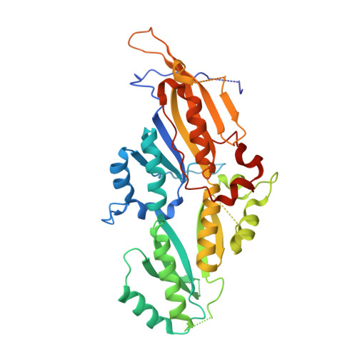
Four KH domains of the C. elegans Bicaudal-C ortholog GLD-3 form a globular structural platform.

Nakel, K.Hartung, S.A.Bonneau, F.Eckmann, C.R.Conti, E.

(2010) RNA 16: 2058-2067

  • DOI: https://doi.org/10.1261/rna.2315010
  • Primary Citation Related Structures: 
    3N89

  • PubMed Abstract: 

    Caenorhabditis elegans GLD-3 is a five K homology (KH) domain-containing protein involved in the translational control of germline-specific mRNAs during embryogenesis. GLD-3 interacts with the cytoplasmic poly(A)-polymerase GLD-2. The two proteins cooperate to recognize target mRNAs and convert them into a polyadenylated, translationally active state. We report the 2.8-Å-resolution crystal structure of a proteolytically stable fragment encompassing the KH2, KH3, KH4, and KH5 domains of C. elegans GLD-3. The structure reveals that the four tandem KH domains are organized into a globular structural unit. The domains are involved in extensive side-by-side interactions, similar to those observed in previous structures of dimeric KH domains, as well as head-to-toe interactions. Small-angle X-ray scattering reconstructions show that the N-terminal KH domain (KH1) forms a thumb-like protrusion on the KH2-KH5 unit. Although KH domains are putative RNA-binding modules, the KH region of GLD-3 is unable in isolation to cross-link RNA. Instead, the KH1 domain mediates the direct interaction with the poly(A)-polymerase GLD-2, pointing to a function of the KH region as a protein-protein interaction platform.


  • Organizational Affiliation
    • Department of Structural Cell Biology, Max-Planck-Institute of Biochemistry, D-82152 Martinsried, Germany.

Macromolecule Content 

  • Total Structure Weight: 85.75 kDa 
  • Atom Count: 5,317 
  • Modeled Residue Count: 656 
  • Deposited Residue Count: 752 
  • Unique protein chains: 1

Macromolecules

Find similar proteins by:|  3D Structure
Entity ID: 1
MoleculeChains  Sequence LengthOrganismDetailsImage
Defective in germ line development protein 3, isoform a
A, B
376Caenorhabditis elegansMutation(s): 0 
Gene Names: gld-3T07F8.3
UniProt
Find proteins for Q95ZK7 (Caenorhabditis elegans)
Explore Q95ZK7 
Go to UniProtKB:  Q95ZK7
Entity Groups
Sequence Clusters30% Identity50% Identity70% Identity90% Identity95% Identity100% Identity
UniProt GroupQ95ZK7
Sequence Annotations
Expand
Reference Sequence

Experimental Data & Validation

Experimental Data

  • Method: X-RAY DIFFRACTION
  • Resolution: 2.79 Å
  • R-Value Free:  0.276 (Depositor), 0.275 (DCC) 
  • R-Value Work:  0.259 (Depositor), 0.258 (DCC) 
Space Group: C 1 2 1
Unit Cell:
Length ( Å )Angle ( ˚ )
a = 159.504α = 90
b = 102.241β = 119.32
c = 83.608γ = 90
Software Package:
Software NamePurpose
CNSrefinement
PDB_EXTRACTdata extraction
XDSdata scaling
XDSdata reduction
XSCALEdata scaling
SHARPphasing

Structure Validation

View Full Validation Report


Entry History 

Deposition Data

Revision History  (Full details and data files)

  • Version 1.0: 2010-09-22
    Type: Initial release
  • Version 1.1: 2011-07-13
    Changes: Version format compliance
  • Version 1.2: 2024-02-21
    Changes: Data collection, Database references