3MTA

Glycogen phosphorylase complexed with 3-bromobenzaldehyde-4-(beta-D-glucopyranosyl)-thiosemicarbazone


Experimental Data Snapshot

  • Method: X-RAY DIFFRACTION
  • Resolution: 2.23 Å
  • R-Value Free: 0.233 
  • R-Value Work: 0.196 
  • R-Value Observed: 0.198 

wwPDB Validation   3D Report Full Report


Ligand Structure Quality Assessment 


This is version 1.5 of the entry. See complete history


Literature

The binding of beta-D-glucopyranosyl-thiosemicarbazone derivatives to glycogen phosphorylase: a new class of inhibitors

Alexacou, K.M.Tenchiu Deleanu, A.C.Chrysina, E.D.Charavgi, M.D.Kostas, I.D.Zographos, S.E.Oikonomakos, N.G.Leonidas, D.D.

(2010) Bioorg Med Chem 18: 7911-7922

  • DOI: https://doi.org/10.1016/j.bmc.2010.09.039
  • Primary Citation of Related Structures:  
    3MQF, 3MRT, 3MRV, 3MRX, 3MS2, 3MS4, 3MS7, 3MSC, 3MT7, 3MT8, 3MT9, 3MTA, 3MTB, 3MTD, 3NC4

  • PubMed Abstract: 

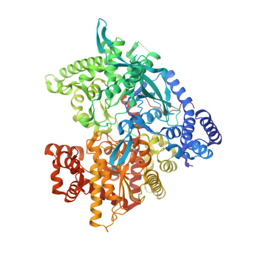
    Glycogen phosphorylase (GP) is a promising target for the treatment of type 2 diabetes. In the process of structure based drug design for GP, a group of 15 aromatic aldehyde 4-(β-d-glucopyranosyl)thiosemicarbazones have been synthesized and evaluated as inhibitors of rabbit muscle glycogen phosphorylase b (GPb) by kinetic studies. These compounds are competitive inhibitors of GPb with respect to α-d-glucose-1-phosphate with IC(50) values ranging from 5.7 to 524.3μM. In order to elucidate the structural basis of their inhibition, the crystal structures of these compounds in complex with GPb at 1.95-2.23Å resolution were determined. The complex structures reveal that the inhibitors are accommodated at the catalytic site with the glucopyranosyl moiety at approximately the same position as α-d-glucose and stabilize the T conformation of the 280s loop. The thiosemicarbazone part of the studied glucosyl thiosemicarbazones possess a moiety derived from substituted benzaldehydes with NO(2), F, Cl, Br, OH, OMe, CF(3), or Me at the ortho-, meta- or para-position of the aromatic ring as well as a moiety derived from 4-pyridinecarboxaldehyde. These fit tightly into the β-pocket, a side channel from the catalytic site with no access to the bulk solvent. The differences in their inhibitory potency can be interpreted in terms of variations in the interactions of the aldehyde-derived moiety with protein residues in the β-pocket. In addition, 14 out of the 15 studied inhibitors were found bound at the new allosteric site of the enzyme.


  • Organizational Affiliation

    Institute of Organic and Pharmaceutical Chemistry, National Hellenic Research Foundation, 48 Vassileos Constantinou Avenue, 11635 Athens, Greece.


Macromolecules
Find similar proteins by:  (by identity cutoff)  |  3D Structure
Entity ID: 1
MoleculeChains Sequence LengthOrganismDetailsImage
Glycogen phosphorylase, muscle form842Oryctolagus cuniculusMutation(s): 0 
EC: 2.4.1.1
UniProt
Find proteins for P00489 (Oryctolagus cuniculus)
Explore P00489 
Go to UniProtKB:  P00489
Entity Groups  
Sequence Clusters30% Identity50% Identity70% Identity90% Identity95% Identity100% Identity
UniProt GroupP00489
Sequence Annotations
Expand
  • Reference Sequence
Small Molecules
Ligands 1 Unique
IDChains Name / Formula / InChI Key2D Diagram3D Interactions
22O
Query on 22O

Download Ideal Coordinates CCD File 
B [auth A],
C [auth A]
N-({(2E)-2-[(3-bromophenyl)methylidene]hydrazino}carbonothioyl)-beta-D-glucopyranosylamine
C14 H18 Br N3 O5 S
PGBHKZPXJWINSR-QCOQDYPWSA-N
Modified Residues  1 Unique
IDChains TypeFormula2D DiagramParent
LLP
Query on LLP
A
L-PEPTIDE LINKINGC14 H22 N3 O7 PLYS
Binding Affinity Annotations 
IDSourceBinding Affinity
22O PDBBind:  3MTA IC50: 5.04e+4 (nM) from 1 assay(s)
Binding MOAD:  3MTA IC50: 5.04e+4 (nM) from 1 assay(s)
Experimental Data & Validation

Experimental Data

  • Method: X-RAY DIFFRACTION
  • Resolution: 2.23 Å
  • R-Value Free: 0.233 
  • R-Value Work: 0.196 
  • R-Value Observed: 0.198 
  • Space Group: P 43 21 2
Unit Cell:
Length ( Å )Angle ( ˚ )
a = 128.916α = 90
b = 128.916β = 90
c = 116.782γ = 90
Software Package:
Software NamePurpose
REFMACrefinement
CNSrefinement
HKL-3000data collection
DENZOdata reduction
SCALEPACKdata scaling
CNSphasing

Structure Validation

View Full Validation Report


Ligand Structure Quality Assessment 


Entry History 

Deposition Data

Revision History  (Full details and data files)

  • Version 1.0: 2011-01-05
    Type: Initial release
  • Version 1.1: 2011-07-13
    Changes: Version format compliance
  • Version 1.2: 2019-07-17
    Changes: Advisory, Data collection, Derived calculations, Refinement description
  • Version 1.3: 2020-07-29
    Type: Remediation
    Reason: Carbohydrate remediation
    Changes: Data collection, Derived calculations
  • Version 1.4: 2023-09-06
    Changes: Data collection, Database references, Refinement description, Structure summary
  • Version 1.5: 2023-11-22
    Changes: Data collection