Crystal structure and mutational analysis of aminoacylhistidine dipeptidase from vibrio alginolyticus reveal a new architecture of M20 metallopeptidases
Chang, C.-Y., Hsieh, Y.-C., Wang, T.-Y., Chen, Y.-C., Wang, Y.-K., Chiang, T.-W., Chen, Y.-J., Chang, C.-H., Chen, C.-J., Wu, T.-K.(2010) J Biological Chem 285: 39500-39510
- PubMed: 20819954 Search on PubMedSearch on PubMed Central
- DOI: https://doi.org/10.1074/jbc.M110.139683
- Primary Citation Related Structures:
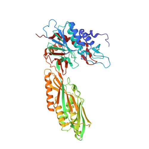
3MRU - PubMed Abstract:
Aminoacylhistidine dipeptidases (PepD, EC 3.4.13.3) belong to the family of M20 metallopeptidases from the metallopeptidase H clan that catalyze a broad range of dipeptide and tripeptide substrates, including L-carnosine and L-homocarnosine. Homocarnosine has been suggested as a precursor for the neurotransmitter γ-aminobutyric acid (GABA) and may mediate the antiseizure effects of GABAergic therapies. Here, we report the crystal structure of PepD from Vibrio alginolyticus and the results of mutational analysis of substrate-binding residues in the C-terminal as well as substrate specificity of the PepD catalytic domain-alone truncated protein PepD(CAT). The structure of PepD was found to exist as a homodimer, in which each monomer comprises a catalytic domain containing two zinc ions at the active site center for its hydrolytic function and a lid domain utilizing hydrogen bonds between helices to form the dimer interface. Although the PepD is structurally similar to PepV, which exists as a monomer, putative substrate-binding residues reside in different topological regions of the polypeptide chain. In addition, the lid domain of the PepD contains an "extra" domain not observed in related M20 family metallopeptidases with a dimeric structure. Mutational assays confirmed both the putative di-zinc allocations and the architecture of substrate recognition. In addition, the catalytic domain-alone truncated PepD(CAT) exhibited substrate specificity to l-homocarnosine compared with that of the wild-type PepD, indicating a potential value in applications of PepD(CAT) for GABAergic therapies or neuroprotection.
- Department of Biological Science and Technology, National Chiao Tung University, Hsinchu 30010, Taiwan.
Organizational Affiliation: