3MQ1

Crystal Structure of Dust Mite Allergen Der p 5


Experimental Data Snapshot

  • Method: X-RAY DIFFRACTION
  • Resolution: 2.80 Å
  • R-Value Free: 0.279 
  • R-Value Work: 0.226 
  • R-Value Observed: 0.229 

wwPDB Validation   3D Report Full Report


This is version 1.2 of the entry. See complete history


Literature

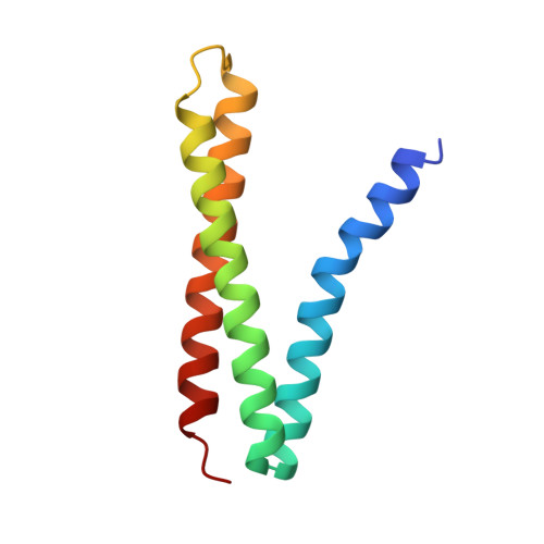
Der p 5 crystal structure provides insight into the group 5 dust mite allergens.

Mueller, G.A.Gosavi, R.A.Krahn, J.M.Edwards, L.L.Cuneo, M.J.Glesner, J.Pomes, A.Chapman, M.D.London, R.E.Pedersen, L.C.

(2010) J Biol Chem 285: 25394-25401

  • DOI: https://doi.org/10.1074/jbc.M110.128306
  • Primary Citation of Related Structures:  
    3MQ1

  • PubMed Abstract: 

    Group 5 allergens from house dust mites elicit strong IgE antibody binding in mite-allergic patients. The structure of Der p 5 was determined by x-ray crystallography to better understand the IgE epitopes, to investigate the biologic function in mites, and to compare with the conflicting published Blo t 5 structures, designated 2JMH and 2JRK in the Protein Data Bank. Der p 5 is a three-helical bundle similar to Blo t 5, but the interactions of the helices are more similar to 2JMH than 2JRK. The crystallographic asymmetric unit contains three dimers of Der p 5 that are not exactly alike. Solution scattering techniques were used to assess the multimeric state of Der p 5 in vitro and showed that the predominant state was monomeric, similar to Blo t 5, but larger multimeric species are also present. In the crystal, the formation of the Der p 5 dimer creates a large hydrophobic cavity of approximately 3000 A(3) that could be a ligand-binding site. Many allergens are known to bind hydrophobic ligands, which are thought to stimulate the innate immune system and have adjuvant-like effects on IgE-mediated inflammatory responses.


  • Organizational Affiliation

    Laboratory of Structural Biology, National Institutes of Health, Research Triangle Park, North Carolina 27709, USA. mueller3@niehs.nih.gov


Macromolecules
Find similar proteins by:  (by identity cutoff)  |  3D Structure
Entity ID: 1
MoleculeChains Sequence LengthOrganismDetailsImage
Mite allergen Der p 5
A, B, C, D, E
A, B, C, D, E, F
103Dermatophagoides pteronyssinusMutation(s): 0 
Gene Names: DERP5
UniProt
Find proteins for P14004 (Dermatophagoides pteronyssinus)
Explore P14004 
Go to UniProtKB:  P14004
Entity Groups  
Sequence Clusters30% Identity50% Identity70% Identity90% Identity95% Identity100% Identity
UniProt GroupP14004
Sequence Annotations
Expand
  • Reference Sequence
Small Molecules
Ligands 3 Unique
IDChains Name / Formula / InChI Key2D Diagram3D Interactions
MRD
Query on MRD

Download Ideal Coordinates CCD File 
BA [auth C]
CA [auth C]
DA [auth C]
EA [auth C]
H [auth A]
BA [auth C],
CA [auth C],
DA [auth C],
EA [auth C],
H [auth A],
JA [auth E],
K [auth A],
KA [auth E],
LA [auth E],
MA [auth E],
NA [auth F],
OA [auth F],
P [auth A],
R [auth A],
T [auth B],
V [auth B],
X [auth B]
(4R)-2-METHYLPENTANE-2,4-DIOL
C6 H14 O2
SVTBMSDMJJWYQN-RXMQYKEDSA-N
MPD
Query on MPD

Download Ideal Coordinates CCD File 
AA [auth C]
FA [auth C]
G [auth A]
GA [auth D]
HA [auth D]
AA [auth C],
FA [auth C],
G [auth A],
GA [auth D],
HA [auth D],
I [auth A],
IA [auth D],
J [auth A],
L [auth A],
M [auth A],
N [auth A],
O [auth A],
Q [auth A],
S [auth B],
W [auth B],
Y [auth B],
Z [auth C]
(4S)-2-METHYL-2,4-PENTANEDIOL
C6 H14 O2
SVTBMSDMJJWYQN-YFKPBYRVSA-N
PO4
Query on PO4

Download Ideal Coordinates CCD File 
U [auth B]PHOSPHATE ION
O4 P
NBIIXXVUZAFLBC-UHFFFAOYSA-K
Experimental Data & Validation

Experimental Data

  • Method: X-RAY DIFFRACTION
  • Resolution: 2.80 Å
  • R-Value Free: 0.279 
  • R-Value Work: 0.226 
  • R-Value Observed: 0.229 
  • Space Group: P 21 21 21
Unit Cell:
Length ( Å )Angle ( ˚ )
a = 46.78α = 90
b = 96.57β = 90
c = 167.89γ = 90
Software Package:
Software NamePurpose
StructureStudiodata collection
PHENIXmodel building
PHENIXrefinement
HKL-2000data reduction
HKL-2000data scaling
PHENIXphasing

Structure Validation

View Full Validation Report


Entry History 

Deposition Data

Revision History  (Full details and data files)

  • Version 1.0: 2010-06-02
    Type: Initial release
  • Version 1.1: 2011-07-13
    Changes: Version format compliance
  • Version 1.2: 2024-02-21
    Changes: Data collection, Database references, Derived calculations