3MI2

Crystal structure of human orotidine-5'-monophosphate decarboxylase complexed with pyrazofurin monophosphate


Experimental Data Snapshot

  • Method: X-RAY DIFFRACTION
  • Resolution: 1.20 Å
  • R-Value Free: 0.192 
  • R-Value Work: 0.171 
  • R-Value Observed: 0.172 

wwPDB Validation   3D Report Full Report


Ligand Structure Quality Assessment 


This is version 1.3 of the entry. See complete history


Literature

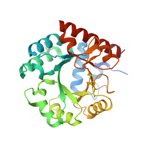
Structural determinants for the inhibitory ligands of orotidine-5'-monophosphate decarboxylase.

Meza-Avina, M.E.Wei, L.Liu, Y.Poduch, E.Bello, A.M.Mishra, R.K.Pai, E.F.Kotra, L.P.

(2010) Bioorg Med Chem 18: 4032-4041

  • DOI: https://doi.org/10.1016/j.bmc.2010.04.017
  • Primary Citation of Related Structures:  
    3MI2

  • PubMed Abstract: 

    In recent years, orotidine-5'-monophosphate decarboxylase (ODCase) has gained renewed attention as a drug target. As a part of continuing efforts to design novel inhibitors of ODCase, we undertook a comprehensive study of potent, structurally diverse ligands of ODCase and analyzed their structural interactions in the active site of ODCase. These ligands comprise of pyrazole or pyrimidine nucleotides including the mononucleotide derivatives of pyrazofurin, barbiturate ribonucleoside, and 5-cyanouridine, as well as, in a computational approach, 1,4-dihydropyridine-based non-nucleoside inhibitors such as nifedipine and nimodipine. All these ligands bind in the active site of ODCase exhibiting distinct interactions paving the way to design novel inhibitors against this interesting enzyme. We propose an empirical model for the ligand structure for rational modifications in new drug design and potentially new lead structures.


  • Organizational Affiliation

    Division of Cell & Molecular Biology, University Health Network, Toronto, ON, Canada.


Macromolecules
Find similar proteins by:  (by identity cutoff)  |  3D Structure
Entity ID: 1
MoleculeChains Sequence LengthOrganismDetailsImage
Uridine 5'-monophosphate synthase
A, B
279Homo sapiensMutation(s): 0 
Gene Names: gi|13960142OK/SW-cl.21UMPS
EC: 4.1.1.23
UniProt & NIH Common Fund Data Resources
Find proteins for P11172 (Homo sapiens)
Explore P11172 
Go to UniProtKB:  P11172
PHAROS:  P11172
GTEx:  ENSG00000114491 
Entity Groups  
Sequence Clusters30% Identity50% Identity70% Identity90% Identity95% Identity100% Identity
UniProt GroupP11172
Sequence Annotations
Expand
  • Reference Sequence
Small Molecules
Ligands 1 Unique
IDChains Name / Formula / InChI Key2D Diagram3D Interactions
PFU
Query on PFU

Download Ideal Coordinates CCD File 
C [auth A],
D [auth B]
(1S)-1,4-anhydro-1-(5-carbamoyl-4-hydroxy-1H-pyrazol-3-yl)-5-O-phosphono-D-ribitol
C9 H14 N3 O9 P
CUHDHRMGDRLFLH-FLLFQEBCSA-N
Modified Residues  1 Unique
IDChains TypeFormula2D DiagramParent
CSD
Query on CSD
A, B
L-PEPTIDE LINKINGC3 H7 N O4 SCYS
Binding Affinity Annotations 
IDSourceBinding Affinity
PFU BindingDB:  3MI2 Ki: 17 (nM) from 1 assay(s)
PDBBind:  3MI2 Ki: 17 (nM) from 1 assay(s)
Binding MOAD:  3MI2 Ki: 790 (nM) from 1 assay(s)
Experimental Data & Validation

Experimental Data

  • Method: X-RAY DIFFRACTION
  • Resolution: 1.20 Å
  • R-Value Free: 0.192 
  • R-Value Work: 0.171 
  • R-Value Observed: 0.172 
  • Space Group: P 1 21 1
Unit Cell:
Length ( Å )Angle ( ˚ )
a = 70.485α = 90
b = 62.109β = 111.45
c = 70.499γ = 90
Software Package:
Software NamePurpose
MxDCdata collection
MOLREPphasing
REFMACrefinement
Cootmodel building
HKL-2000data reduction
HKL-2000data scaling

Structure Validation

View Full Validation Report


Ligand Structure Quality Assessment 


Entry History 

Deposition Data

Revision History  (Full details and data files)

  • Version 1.0: 2010-05-26
    Type: Initial release
  • Version 1.1: 2011-07-13
    Changes: Version format compliance
  • Version 1.2: 2014-11-12
    Changes: Structure summary
  • Version 1.3: 2023-09-06
    Changes: Data collection, Database references, Derived calculations, Refinement description