Crystal structures, dynamics and functional implications of molybdenum-cofactor biosynthesis protein MogA from two thermophilic organisms
Kanaujia, S.P., Jeyakanthan, J., Shinkai, A., Kuramitsu, S., Yokoyama, S., Sekar, K.(2011) Acta Crystallogr Sect F Struct Biol Cryst Commun 67: 2-16
- PubMed: 21206014 Search on PubMedSearch on PubMed Central
- DOI: https://doi.org/10.1107/S1744309110035037
- Primary Citation Related Structures:
3MCH, 3MCI, 3MCJ - PubMed Abstract:
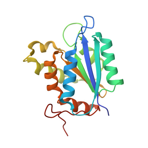
Molybdenum-cofactor (Moco) biosynthesis is an evolutionarily conserved pathway in almost all kingdoms of life, including humans. Two proteins, MogA and MoeA, catalyze the last step of this pathway in bacteria, whereas a single two-domain protein carries out catalysis in eukaryotes. Here, three crystal structures of the Moco-biosynthesis protein MogA from the two thermophilic organisms Thermus thermophilus (TtMogA; 1.64 Å resolution, space group P2(1)) and Aquifex aeolicus (AaMogA; 1.70 Å resolution, space group P2(1) and 1.90 Å resolution, space group P1) have been determined. The functional roles and the residues involved in oligomerization of the protein molecules have been identified based on a comparative analysis of these structures with those of homologous proteins. Furthermore, functional roles have been proposed for the N- and C-terminal residues. In addition, a possible protein-protein complex of MogA and MoeA has been proposed and the residues involved in protein-protein interactions are discussed. Several invariant water molecules and those present at the subunit interfaces have been identified and their possible structural and/or functional roles are described in brief. In addition, molecular-dynamics and docking studies with several small molecules (including the substrate and the product) have been carried out in order to estimate their binding affinities towards AaMogA and TtMogA. The results obtained are further compared with those obtained for homologous eukaryotic proteins.
- Bioinformatics Centre, Centre of Excellence in Structural Biology and Bio-computing, Indian Institute of Science, Bangalore 560 012, India.
Organizational Affiliation: