The structure and evolution of the murine inhibitor of carbonic anhydrase: A member of the transferrin superfamily.
Eckenroth, B.E., Mason, A.B., McDevitt, M.E., Lambert, L.A., Everse, S.J.(2010) Protein Sci 19: 1616-1626
- PubMed: 20572014 Search on PubMedSearch on PubMed Central
- DOI: https://doi.org/10.1002/pro.439
- Primary Citation Related Structures:
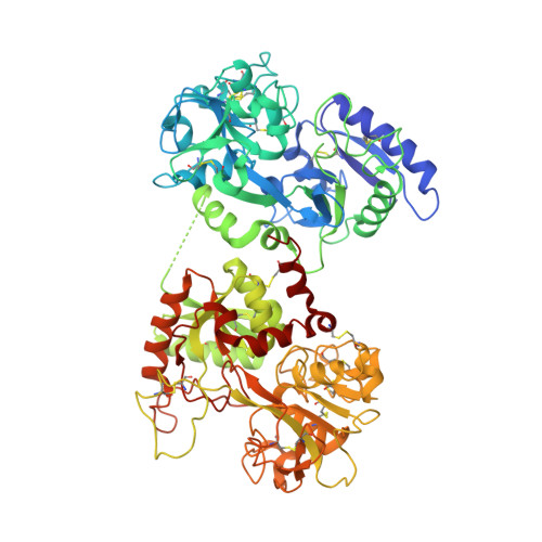
3MC2 - PubMed Abstract:
The original signature of the transferrin (TF) family of proteins was the ability to bind ferric iron with high affinity in the cleft of each of two homologous lobes. However, in recent years, new family members that do not bind iron have been discovered. One new member is the inhibitor of carbonic anhydrase (ICA), which as its name indicates, binds to and strongly inhibits certain isoforms of carbonic anhydrase. Recently, mouse ICA has been expressed as a recombinant protein in a mammalian cell system. Here, we describe the 2.4 Å structure of mouse ICA from a pseudomerohedral twinned crystal. As predicted, the structure is bilobal, comprised of two α-β domains per lobe typical of the other family members. As with all but insect TFs, the structure includes the unusual reverse γ-turn in each lobe. The structure is consistent with the fact that introduction of two mutations in the N-lobe of murine ICA (mICA) (W124R and S188Y) allowed it to bind iron with high affinity. Unexpectedly, both lobes of the mICA were found in the closed conformation usually associated with presence of iron in the cleft, and making the structure most similar to diferric pig TF. Two new ICA family members (guinea pig and horse) were identified from genomic sequences and used in evolutionary comparisons. Additionally, a comparison of selection pressure (dN/dS) on functional residues reveals some interesting insights into the evolution of the TF family including that the N-lobe of lactoferrin may be in the process of eliminating its iron binding function.
- Department of Biochemistry, University of Vermont, Burlington, Vermont 05405, USA.
Organizational Affiliation: