3MBL | pdb_00003mbl

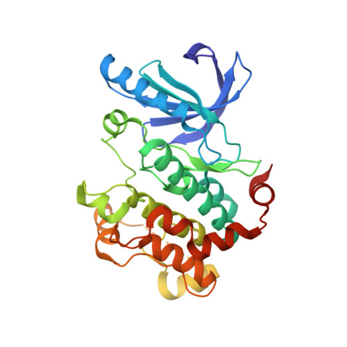
Crystal Structure of the human mitogen-activated protein kinase kinase 1 (MEK 1) in complex with ligand and MgADP


Experimental Data Snapshot

  • Method: X-RAY DIFFRACTION
  • Resolution: 2.60 Å
  • R-Value Free: 
    0.215 (Depositor), 0.213 (DCC) 
  • R-Value Work: 
    0.166 (Depositor), 0.162 (DCC) 
  • R-Value Observed: 
    0.169 (Depositor) 

wwPDB Validation 3D Report Full Report

Validation slider image for 3MBL

Ligand Structure Quality Assessment 


This is version 1.2 of the entry. See complete history. 

Literature

Structure-based design and synthesis of pyrrole derivatives as MEK inhibitors.

Wallace, M.B., Adams, M.E., Kanouni, T., Mol, C.D., Dougan, D.R., Feher, V.A., O'Connell, S.M., Shi, L., Halkowycz, P., Dong, Q.

(2010) Bioorg Med Chem Lett 20: 4156-4158

  • DOI: https://doi.org/10.1016/j.bmcl.2010.05.058
  • Primary Citation Related Structures: 
    3MBL

  • PubMed Abstract: 

    A novel series of pyrrole inhibitors of MEK kinase has been developed using structure-based drug design. Optimization of the series led to the identification of potent inhibitors with good pharmaceutical properties.


  • Organizational Affiliation: 
    • Takeda San Diego, 10410 Science Center Drive, San Diego, CA 92121, United States. michael.wallace@takedasd.com

Macromolecule Content 

  • Total Structure Weight: 37.55 kDa 
  • Atom Count: 2,420 
  • Modeled Residue Count: 290 
  • Deposited Residue Count: 328 
  • Unique protein chains: 1

Macromolecules

Find similar proteins by:|  3D Structure
Entity ID: 1
MoleculeChains  Sequence LengthOrganismDetailsImage
Dual specificity mitogen-activated protein kinase kinase 1328Homo sapiensMutation(s): 0 
Gene Names: MAP2K1, MEK 1, MEK1, PRKMK1
EC: 2.7.12.2
UniProt & NIH Common Fund Data Resources
Find proteins for Q02750 (Homo sapiens)
Explore Q02750 
Go to UniProtKB:  Q02750
PHAROS:  Q02750
GTEx:  ENSG00000169032 
Entity Groups
Sequence Clusters30% Identity50% Identity70% Identity90% Identity95% Identity100% Identity
UniProt GroupQ02750
Sequence Annotations
Expand
Reference Sequence

Small Molecules

Ligands 3 Unique
IDChains Name / Formula / InChI Key2D Diagram3D Interactions
LSG

Query on LSG


Download:Ideal Coordinates CCD File
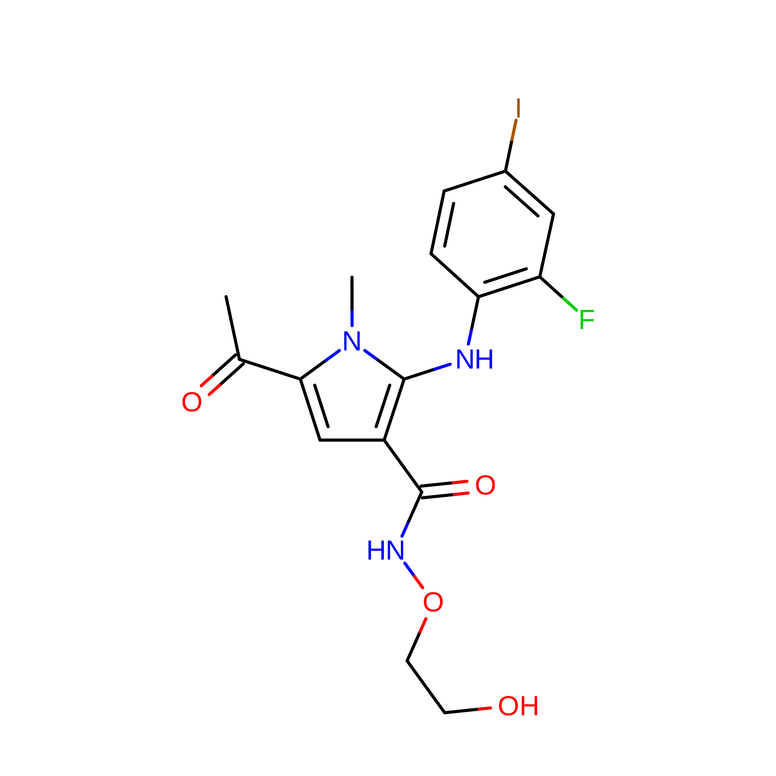
D [auth A]5-acetyl-2-[(2-fluoro-4-iodophenyl)amino]-N-(2-hydroxyethoxy)-1-methyl-1H-pyrrole-3-carboxamide
C16 H17 F I N3 O4
WRHOGNMBUWCIAC-UHFFFAOYSA-N
ADP

Query on ADP


Download:Ideal Coordinates CCD File
B [auth A]ADENOSINE-5'-DIPHOSPHATE
C10 H15 N5 O10 P2
XTWYTFMLZFPYCI-KQYNXXCUSA-N
MG

Query on MG


Download:Ideal Coordinates CCD File
C [auth A]MAGNESIUM ION
Mg
JLVVSXFLKOJNIY-UHFFFAOYSA-N
Binding Affinity Annotations 
IDSourceBinding Affinity
LSG BindingDB:  3MBL IC50: 18 (nM) from 1 assay(s)

Experimental Data & Validation

Experimental Data

  • Method: X-RAY DIFFRACTION
  • Resolution: 2.60 Å
  • R-Value Free:  0.215 (Depositor), 0.213 (DCC) 
  • R-Value Work:  0.166 (Depositor), 0.162 (DCC) 
  • R-Value Observed: 0.169 (Depositor) 
Space Group: P 62
Unit Cell:
Length ( Å )Angle ( ˚ )
a = 81.982α = 90
b = 81.982β = 90
c = 129.928γ = 120
Software Package:
Software NamePurpose
HKL-2000data collection
MOLREPphasing
REFMACrefinement
HKL-2000data reduction
HKL-2000data scaling

Structure Validation

View Full Validation Report


Ligand Structure Quality Assessment 


Entry History 

Deposition Data

Revision History  (Full details and data files)

  • Version 1.0: 2010-07-28
    Type: Initial release
  • Version 1.1: 2011-07-13
    Changes: Version format compliance
  • Version 1.2: 2024-02-21
    Changes: Data collection, Database references, Derived calculations