3M9C

Crystal structure of the membrane domain of respiratory complex I from Escherichia coli


Experimental Data Snapshot

  • Method: X-RAY DIFFRACTION
  • Resolution: 3.90 Å

wwPDB Validation   3D Report Full Report


This is version 1.3 of the entry. See complete history


Literature

The architecture of respiratory complex I

Efremov, R.G.Baradaran, R.Sazanov, L.A.

(2010) Nature 465: 441-445

  • DOI: https://doi.org/10.1038/nature09066
  • Primary Citation of Related Structures:  
    3M9C, 3M9S

  • PubMed Abstract: 

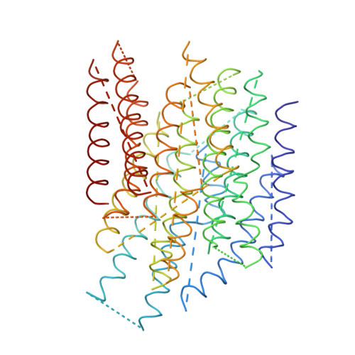
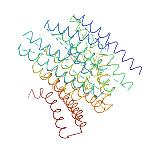
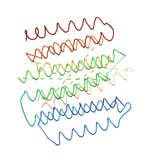
    Complex I is the first enzyme of the respiratory chain and has a central role in cellular energy production, coupling electron transfer between NADH and quinone to proton translocation by an unknown mechanism. Dysfunction of complex I has been implicated in many human neurodegenerative diseases. We have determined the structure of its hydrophilic domain previously. Here, we report the alpha-helical structure of the membrane domain of complex I from Escherichia coli at 3.9 A resolution. The antiporter-like subunits NuoL/M/N each contain 14 conserved transmembrane (TM) helices. Two of them are discontinuous, as in some transporters. Unexpectedly, subunit NuoL also contains a 110-A long amphipathic alpha-helix, spanning almost the entire length of the domain. Furthermore, we have determined the structure of the entire complex I from Thermus thermophilus at 4.5 A resolution. The L-shaped assembly consists of the alpha-helical model for the membrane domain, with 63 TM helices, and the known structure of the hydrophilic domain. The architecture of the complex provides strong clues about the coupling mechanism: the conformational changes at the interface of the two main domains may drive the long amphipathic alpha-helix of NuoL in a piston-like motion, tilting nearby discontinuous TM helices, resulting in proton translocation.


  • Organizational Affiliation

    Medical Research Council Mitochondrial Biology Unit, Wellcome Trust/MRC Building, Hills Road, Cambridge CB2 0XY, UK.


Macromolecules
Find similar proteins by:  (by identity cutoff)  |  3D Structure
Entity ID: 1
MoleculeChains Sequence LengthOrganismDetailsImage
NADH-quinone oxidoreductase subunit NuoLA [auth L]474Escherichia coliMutation(s): 0 
EC: 1.6.5.3
Sequence Annotations
Expand
  • Reference Sequence
Find similar proteins by:  (by identity cutoff)  |  3D Structure
Entity ID: 2
MoleculeChains Sequence LengthOrganismDetailsImage
NADH-quinone oxidoreductase subunit NuoMB [auth M]391Escherichia coliMutation(s): 0 
EC: 1.6.5.3
Sequence Annotations
Expand
  • Reference Sequence
Find similar proteins by:  (by identity cutoff)  |  3D Structure
Entity ID: 3
MoleculeChains Sequence LengthOrganismDetailsImage
NADH-quinone oxidoreductase subunit NuoNC [auth N]378Escherichia coliMutation(s): 0 
EC: 1.6.5.3
Sequence Annotations
Expand
  • Reference Sequence
Find similar proteins by:  (by identity cutoff)  |  3D Structure
Entity ID: 4
MoleculeChains Sequence LengthOrganismDetailsImage
NADH-quinone oxidoreductase subunits NuoA,J and KD [auth R]281Escherichia coliMutation(s): 0 
EC: 1.6.5.3
Sequence Annotations
Expand
  • Reference Sequence
Experimental Data & Validation

Experimental Data

  • Method: X-RAY DIFFRACTION
  • Resolution: 3.90 Å
  • Space Group: P 21 21 21
Unit Cell:
Length ( Å )Angle ( ˚ )
a = 114.06α = 90
b = 182.17β = 90
c = 185.6γ = 90
Software Package:
Software NamePurpose
MOSFLMdata reduction
PHENIXmodel building
Omodel building
Cootmodel building
SCALAdata scaling
PHENIXphasing

Structure Validation

View Full Validation Report


Entry History 

Deposition Data

Revision History  (Full details and data files)

  • Version 1.0: 2010-05-26
    Type: Initial release
  • Version 1.1: 2011-07-13
    Changes: Version format compliance
  • Version 1.2: 2015-01-21
    Changes: Database references
  • Version 1.3: 2024-02-21
    Changes: Database references