Ultrahigh (0.93A) resolution structure of manganese peroxidase from Phanerochaete chrysosporium: implications for the catalytic mechanism.
Sundaramoorthy, M., Gold, M.H., Poulos, T.L.(2010) J Inorg Biochem 104: 683-690
- PubMed: 20356630 Search on PubMedSearch on PubMed Central
- DOI: https://doi.org/10.1016/j.jinorgbio.2010.02.011
- Primary Citation Related Structures:
3M5Q, 3M8M - PubMed Abstract:
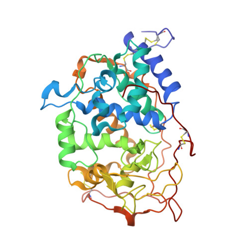
Manganese peroxidase (MnP) is an extracellular heme enzyme produced by the lignin-degrading white-rot fungus Phanerochaete chrysosporium. MnP catalyzes the peroxide-dependent oxidation of Mn(II) to Mn(III). The Mn(III) is released from the enzyme in complex with oxalate, enabling the oxalate-Mn(III) complex to serve as a diffusible redox mediator capable of oxidizing lignin, especially under the mediation of unsaturated fatty acids. One heme propionate and the side chains of Glu35, Glu39 and Asp179 have been identified as Mn(II) ligands in our previous crystal structures of native MnP. In our current work, new 0.93A and 1.05A crystal structures of MnP with and without bound Mn(II), respectively, have been solved. This represents only the sixth structure of a protein of this size at 0.93A resolution. In addition, this is the first structure of a heme peroxidase from a eukaryotic organism at sub-Angstrom resolution. These new structures reveal an ordering/disordering of the C-terminal loop, which is likely required for Mn binding and release. In addition, the catalytic Arg42 residue at the active site, normally thought to function only in the peroxide activation process, also undergoes ordering/disordering that is coupled to a transient H-bond with the Mn ligand, Glu39. Finally, these high-resolution structures also reveal the exact H atoms in several parts of the structure that are relevant to the catalytic mechanism.
- Department of Biochemistry, Vanderbilt University Medical Center, Nashville, TN 37232, United States.
Organizational Affiliation: