3LQF

Crystal structure of the short-chain dehydrogenase Galactitol-Dehydrogenase (GatDH) of Rhodobacter sphaeroides in complex with NAD and erythritol


Experimental Data Snapshot

  • Method: X-RAY DIFFRACTION
  • Resolution: 1.80 Å
  • R-Value Free: 0.223 
  • R-Value Work: 0.184 
  • R-Value Observed: 0.186 

wwPDB Validation   3D Report Full Report


Ligand Structure Quality Assessment 


This is version 1.3 of the entry. See complete history


Literature

Structural insight into substrate differentiation of the sugar-metabolizing enzyme galactitol dehydrogenase from Rhodobacter sphaeroides D.

Carius, Y.Christian, H.Faust, A.Zander, U.Klink, B.U.Kornberger, P.Kohring, G.W.Giffhorn, F.Scheidig, A.J.

(2010) J Biol Chem 285: 20006-20014

  • DOI: https://doi.org/10.1074/jbc.M110.113738
  • Primary Citation of Related Structures:  
    2WDZ, 2WSB, 3LQF

  • PubMed Abstract: 

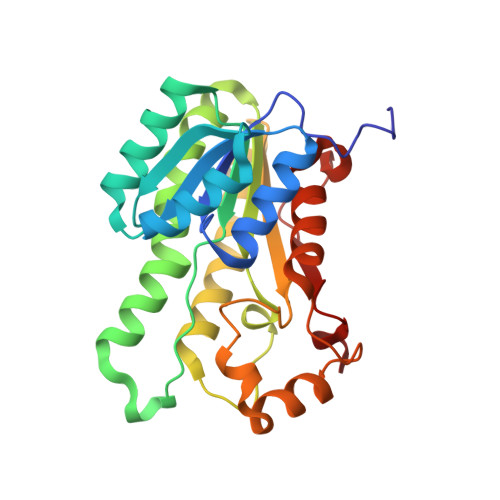
    Galactitol 2-dehydrogenase (GatDH) belongs to the protein superfamily of short-chain dehydrogenases. As an enzyme capable of the stereo- and regioselective modification of carbohydrates, it exhibits a high potential for application in biotechnology as a biocatalyst. We have determined the crystal structure of the binary form of GatDH in complex with its cofactor NAD(H) and of the ternary form in complex with NAD(H) and three different substrates. The active form of GatDH constitutes a homo-tetramer with two magnesium-ion binding sites each formed by two opposing C termini. The catalytic tetrad is formed by Asn(116), Ser(144), Tyr(159), and Lys(163). GatDH structurally aligns well with related members of the short-chain dehydrogenase family. The substrate binding pocket can be divided into two parts of different size and polarity. In the smaller part, the side chains of amino acids Ser(144), Ser(146), and Asn(151) are important determinants for the binding specificity and the orientation of (pro-) chiral compounds. The larger part of the pocket is elongated and flanked by polar and non-polar residues, enabling a rather broad substrate spectrum. The presented structures provide valuable information for a rational design of this enzyme to improve its stability against pH, temperature, or solvent concentration and to optimize product yield in bioreactors.


  • Organizational Affiliation

    Department of Structural Biology, Zoological Institute, Christian-Albrechts-University Kiel, Am Botanischen Garten 1-9, D-24118 Kiel, Germany.


Macromolecules
Find similar proteins by:  (by identity cutoff)  |  3D Structure
Entity ID: 1
MoleculeChains Sequence LengthOrganismDetailsImage
Galactitol dehydrogenase
A, B, C, D
254Cereibacter sphaeroidesMutation(s): 0 
EC: 1.1.1.16
UniProt
Find proteins for C0KTJ6 (Cereibacter sphaeroides)
Explore C0KTJ6 
Go to UniProtKB:  C0KTJ6
Entity Groups  
Sequence Clusters30% Identity50% Identity70% Identity90% Identity95% Identity100% Identity
UniProt GroupC0KTJ6
Sequence Annotations
Expand
  • Reference Sequence
Small Molecules
Ligands 3 Unique
IDChains Name / Formula / InChI Key2D Diagram3D Interactions
NAD
Query on NAD

Download Ideal Coordinates CCD File 
F [auth A],
H [auth B],
J [auth C],
M [auth D]
NICOTINAMIDE-ADENINE-DINUCLEOTIDE
C21 H27 N7 O14 P2
BAWFJGJZGIEFAR-NNYOXOHSSA-N
MRY
Query on MRY

Download Ideal Coordinates CCD File 
G [auth A],
I [auth B],
K [auth C],
N [auth D]
MESO-ERYTHRITOL
C4 H10 O4
UNXHWFMMPAWVPI-ZXZARUISSA-N
MG
Query on MG

Download Ideal Coordinates CCD File 
E [auth A],
L [auth C]
MAGNESIUM ION
Mg
JLVVSXFLKOJNIY-UHFFFAOYSA-N
Experimental Data & Validation

Experimental Data

  • Method: X-RAY DIFFRACTION
  • Resolution: 1.80 Å
  • R-Value Free: 0.223 
  • R-Value Work: 0.184 
  • R-Value Observed: 0.186 
  • Space Group: P 21 21 21
Unit Cell:
Length ( Å )Angle ( ˚ )
a = 63.863α = 90
b = 113.838β = 90
c = 123.234γ = 90
Software Package:
Software NamePurpose
MOLREPphasing
REFMACrefinement
XDSdata reduction
XDSdata scaling

Structure Validation

View Full Validation Report


Ligand Structure Quality Assessment 


Entry History 

Deposition Data

Revision History  (Full details and data files)

  • Version 1.0: 2010-04-21
    Type: Initial release
  • Version 1.1: 2011-07-13
    Changes: Version format compliance
  • Version 1.2: 2024-02-21
    Changes: Data collection, Database references, Derived calculations
  • Version 1.3: 2024-04-03
    Changes: Refinement description