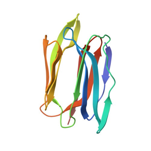
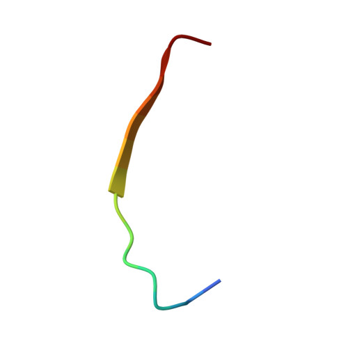
3LLY

Crystal Structure Analysis of Maclura pomifera agglutinin


Experimental Data Snapshot

  • Method: X-RAY DIFFRACTION
  • Resolution: 2.25 Å
  • R-Value Free: 0.263 
  • R-Value Work: 0.217 

Starting Model: experimental
View more details

wwPDB Validation   3D Report Full Report


This is version 1.2 of the entry. See complete history


Literature

Characterization of the secondary binding sites of Maclura pomifera agglutinin by glycan array and crystallographic analyses.

Huang, J.Xu, Z.Wang, D.Ogata, C.M.Palczewski, K.Lee, X.Young, N.M.

(2010) Glycobiology 20: 1643-1653

  • DOI: https://doi.org/10.1093/glycob/cwq118
  • Primary Citation of Related Structures:  
    3LLY, 3LLZ, 3LM1

  • PubMed Abstract: 

    The Maclura pomifera agglutinin (MPA) recognizes the T-antigen disaccharide Galβ1,3GalNAc mainly through interaction of the α-GalNAc moiety with its primary site, but the interactions of the two flanking subsites A and B with aglycones and substituents other than Gal, respectively, are not well understood. We therefore characterized the specificity of MPA in more detail by glycan microarray analysis and determined the crystal structures of MPA without ligand and in complexes with Galβ1,3GalNAc and p-nitrophenyl α-GalNAc. In both sugar complexes, pairs of ligands created inter-tetramer hydrogen-bond bridging networks. While subsite A showed increased affinity for hydrophobic aglycones, it also accommodated several sugar substituents. Notably, a GalNAc-O-tripeptide, a Tn-antigen mimic, showed lower affinity than these compounds in surface plasmon resonance (SPR) experiments. The glycan array data that showed subsite B accepted compounds in which the O3 position of the GalNAc was substituted with various sugars other than Gal, but substitutions at O6 led to inactivity. Additions to the Gal moiety of the disaccharide also had only small effects on reactivity. These results are all compatible with the features seen in the crystal structures.


  • Organizational Affiliation

    Department of Pharmacology, School of Medicine, Case Western Reserve University, 2109 Adelbert Rd, Cleveland, OH 44106, USA.


Macromolecules
Find similar proteins by:  (by identity cutoff)  |  3D Structure
Entity ID: 1
MoleculeChains Sequence LengthOrganismDetailsImage
Agglutinin alpha chain133Maclura pomiferaMutation(s): 0 
UniProt
Find proteins for P18674 (Maclura pomifera)
Explore P18674 
Go to UniProtKB:  P18674
Entity Groups  
Sequence Clusters30% Identity50% Identity70% Identity90% Identity95% Identity100% Identity
UniProt GroupP18674
Sequence Annotations
Expand
  • Reference Sequence

Find similar proteins by:  Sequence   |   3D Structure  

Entity ID: 2
MoleculeChains Sequence LengthOrganismDetailsImage
Agglutinin beta-2 chain16Maclura pomiferaMutation(s): 0 
UniProt
Find proteins for P18676 (Maclura pomifera)
Explore P18676 
Go to UniProtKB:  P18676
Entity Groups  
Sequence Clusters30% Identity50% Identity70% Identity90% Identity95% Identity100% Identity
UniProt GroupP18676
Sequence Annotations
Expand
  • Reference Sequence
Experimental Data & Validation

Experimental Data

  • Method: X-RAY DIFFRACTION
  • Resolution: 2.25 Å
  • R-Value Free: 0.263 
  • R-Value Work: 0.217 
  • Space Group: P 64 2 2
Unit Cell:
Length ( Å )Angle ( ˚ )
a = 61.753α = 90
b = 61.753β = 90
c = 149.537γ = 120
Software Package:
Software NamePurpose
CNSrefinement
PDB_EXTRACTdata extraction
APEXdata collection
HKL-2000data reduction
HKL-2000data scaling
AMoREphasing

Structure Validation

View Full Validation Report


Entry History 

Deposition Data

Revision History  (Full details and data files)

  • Version 1.0: 2010-09-22
    Type: Initial release
  • Version 1.1: 2011-07-13
    Changes: Version format compliance
  • Version 1.2: 2023-09-06
    Changes: Data collection, Database references, Refinement description