Experimental Data Snapshot

  • Method: X-RAY DIFFRACTION
  • Resolution: 2.80 Å
  • R-Value Free: 
    0.238 (Depositor), 0.237 (DCC) 
  • R-Value Work: 
    0.211 (Depositor), 0.215 (DCC) 
  • R-Value Observed: 
    0.212 (Depositor) 

wwPDB Validation 3D Report Full Report

Validation slider image for 3LLM

Ligand Structure Quality Assessment 


This is version 1.2 of the entry. See complete history

Literature

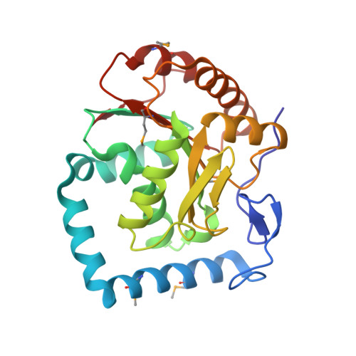
Crystal structure of human RNA helicase A (DHX9): structural basis for unselective nucleotide base binding in a DEAD-box variant protein.

Schutz, P.Wahlberg, E.Karlberg, T.Hammarstrom, M.Collins, R.Flores, A.Schuler, H.

(2010) J Mol Biology 400: 768-782

  • DOI: https://doi.org/10.1016/j.jmb.2010.05.046
  • Primary Citation Related Structures: 
    3LLM

  • PubMed Abstract: 

    RNA helicases of the DExD/H-box superfamily are critically involved in all RNA-related processes. No crystal structures of human DExH-box domains had been determined previously, and their structures were difficult to predict owing to the low level of homology among DExH-motif-containing proteins from diverse species. Here we present the crystal structures of the conserved domain 1 of the DEIH-motif-containing helicase DHX9 and of the DEAD-box helicase DDX20. Both contain a RecA-like core, but DHX9 differs from DEAD-box proteins in the arrangement of secondary structural elements and is more similar to viral helicases such as NS3. The N-terminus of the DHX9 core contains two long alpha-helices that reside on the surface of the core without contributing to nucleotide binding. The RNA-polymerase-II-interacting minimal transactivation domain sequence forms an extended loop structure that resides in a hydrophobic groove on the surface of the DEIH domain. DHX9 lacks base-selective contacts and forms an unspecific but important stacking interaction with the base of the bound nucleotide, and our biochemical analysis confirms that the protein can hydrolyze ATP, guanosine 5'-triphosphate, cytidine 5'-triphosphate, and uridine 5'-triphosphate. Together, these findings allow the localization of functional motifs within the three-dimensional structure of a human DEIH helicase and show how these enzymes can bind nucleotide with high affinity in the absence of a Q-motif.


  • Organizational Affiliation
    • Department of Medical Biochemistry and Biophysics, Karolinska Institutet, Stockholm, Sweden.

Macromolecule Content 

  • Total Structure Weight: 54.72 kDa 
  • Atom Count: 3,773 
  • Modeled Residue Count: 470 
  • Deposited Residue Count: 470 
  • Unique protein chains: 1

Macromolecules

Find similar proteins by:|  3D Structure
Entity ID: 1
MoleculeChains  Sequence LengthOrganismDetailsImage
ATP-dependent RNA helicase A
A, B
235Homo sapiensMutation(s): 0 
Gene Names: DDX9DHX9LKPNDH2
EC: 3.6.1 (PDB Primary Data), 3.6.4.13 (UniProt)
UniProt & NIH Common Fund Data Resources
Find proteins for Q08211 (Homo sapiens)
Explore Q08211 
Go to UniProtKB:  Q08211
PHAROS:  Q08211
GTEx:  ENSG00000135829 
Entity Groups
Sequence Clusters30% Identity50% Identity70% Identity90% Identity95% Identity100% Identity
UniProt GroupQ08211
Sequence Annotations
Expand
Reference Sequence

Small Molecules

Ligands 4 Unique
IDChains Name / Formula / InChI Key2D Diagram3D Interactions
ADP

Query on ADP


Download:Ideal Coordinates CCD File
E [auth A],
G [auth B]
ADENOSINE-5'-DIPHOSPHATE
C10 H15 N5 O10 P2
XTWYTFMLZFPYCI-KQYNXXCUSA-N
CAC

Query on CAC


Download:Ideal Coordinates CCD File
D [auth A]CACODYLATE ION
C2 H6 As O2
OGGXGZAMXPVRFZ-UHFFFAOYSA-M
GOL

Query on GOL


Download:Ideal Coordinates CCD File
H [auth B]GLYCEROL
C3 H8 O3
PEDCQBHIVMGVHV-UHFFFAOYSA-N
MN

Query on MN


Download:Ideal Coordinates CCD File
C [auth A],
F [auth B]
MANGANESE (II) ION
Mn
WAEMQWOKJMHJLA-UHFFFAOYSA-N
Modified Residues  1 Unique
IDChains TypeFormula2D DiagramParent
MSE
Query on MSE
A, B
L-PEPTIDE LINKINGC5 H11 N O2 SeMET

Experimental Data & Validation

Experimental Data

  • Method: X-RAY DIFFRACTION
  • Resolution: 2.80 Å
  • R-Value Free:  0.238 (Depositor), 0.237 (DCC) 
  • R-Value Work:  0.211 (Depositor), 0.215 (DCC) 
  • R-Value Observed: 0.212 (Depositor) 
Space Group: P 32 2 1
Unit Cell:
Length ( Å )Angle ( ˚ )
a = 113.7α = 90
b = 113.7β = 90
c = 141.65γ = 120
Software Package:
Software NamePurpose
SCALAdata scaling
REFMACrefinement
PDB_EXTRACTdata extraction
GDAdata collection
MOSFLMdata reduction
SHARPphasing

Structure Validation

View Full Validation Report


Ligand Structure Quality Assessment 


Entry History 

Revision History  (Full details and data files)

  • Version 1.0: 2010-05-12
    Type: Initial release
  • Version 1.1: 2011-07-13
    Changes: Version format compliance
  • Version 1.2: 2024-11-27
    Changes: Data collection, Database references, Derived calculations, Refinement description, Structure summary