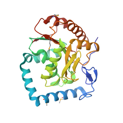
3LLM | pdb_00003llm

Crystal Structure Analysis of a RNA Helicase


Experimental Data Snapshot

  • Method: X-RAY DIFFRACTION
  • Resolution: 2.80 Å
  • R-Value Free: 
    0.238 (Depositor), 0.237 (DCC) 
  • R-Value Work: 
    0.211 (Depositor), 0.215 (DCC) 
  • R-Value Observed: 
    0.212 (Depositor) 

wwPDB Validation   3D Report Full Report


Ligand Structure Quality Assessment 


This is version 1.2 of the entry. See complete history


Literature

Crystal structure of human RNA helicase A (DHX9): structural basis for unselective nucleotide base binding in a DEAD-box variant protein.

Schutz, P.Wahlberg, E.Karlberg, T.Hammarstrom, M.Collins, R.Flores, A.Schuler, H.

(2010) J Mol Biology 400: 768-782

  • DOI: https://doi.org/10.1016/j.jmb.2010.05.046
  • Primary Citation of Related Structures:  
    3LLM

  • PubMed Abstract: 

    RNA helicases of the DExD/H-box superfamily are critically involved in all RNA-related processes. No crystal structures of human DExH-box domains had been determined previously, and their structures were difficult to predict owing to the low level of homology among DExH-motif-containing proteins from diverse species. Here we present the crystal structures of the conserved domain 1 of the DEIH-motif-containing helicase DHX9 and of the DEAD-box helicase DDX20. Both contain a RecA-like core, but DHX9 differs from DEAD-box proteins in the arrangement of secondary structural elements and is more similar to viral helicases such as NS3. The N-terminus of the DHX9 core contains two long alpha-helices that reside on the surface of the core without contributing to nucleotide binding. The RNA-polymerase-II-interacting minimal transactivation domain sequence forms an extended loop structure that resides in a hydrophobic groove on the surface of the DEIH domain. DHX9 lacks base-selective contacts and forms an unspecific but important stacking interaction with the base of the bound nucleotide, and our biochemical analysis confirms that the protein can hydrolyze ATP, guanosine 5'-triphosphate, cytidine 5'-triphosphate, and uridine 5'-triphosphate. Together, these findings allow the localization of functional motifs within the three-dimensional structure of a human DEIH helicase and show how these enzymes can bind nucleotide with high affinity in the absence of a Q-motif.


  • Organizational Affiliation

    Department of Medical Biochemistry and Biophysics, Karolinska Institutet, Stockholm, Sweden.


Macromolecules
Find similar proteins by:  (by identity cutoff)  |  3D Structure
Entity ID: 1
MoleculeChains Sequence LengthOrganismDetailsImage
ATP-dependent RNA helicase A
A, B
235Homo sapiensMutation(s): 0 
Gene Names: DDX9DHX9LKPNDH2
EC: 3.6.1 (PDB Primary Data), 3.6.4.13 (UniProt)
UniProt & NIH Common Fund Data Resources
Find proteins for Q08211 (Homo sapiens)
Explore Q08211 
Go to UniProtKB:  Q08211
PHAROS:  Q08211
GTEx:  ENSG00000135829 
Entity Groups  
Sequence Clusters30% Identity50% Identity70% Identity90% Identity95% Identity100% Identity
UniProt GroupQ08211
Sequence Annotations
Expand
  • Reference Sequence
Small Molecules
Ligands 4 Unique
IDChains Name / Formula / InChI Key2D Diagram3D Interactions
ADP
Query on ADP

Download Ideal Coordinates CCD File 
E [auth A],
G [auth B]
ADENOSINE-5'-DIPHOSPHATE
C10 H15 N5 O10 P2
XTWYTFMLZFPYCI-KQYNXXCUSA-N
CAC
Query on CAC

Download Ideal Coordinates CCD File 
D [auth A]CACODYLATE ION
C2 H6 As O2
OGGXGZAMXPVRFZ-UHFFFAOYSA-M
GOL
Query on GOL

Download Ideal Coordinates CCD File 
H [auth B]GLYCEROL
C3 H8 O3
PEDCQBHIVMGVHV-UHFFFAOYSA-N
MN
Query on MN

Download Ideal Coordinates CCD File 
C [auth A],
F [auth B]
MANGANESE (II) ION
Mn
WAEMQWOKJMHJLA-UHFFFAOYSA-N
Modified Residues  1 Unique
IDChains TypeFormula2D DiagramParent
MSE
Query on MSE
A, B
L-PEPTIDE LINKINGC5 H11 N O2 SeMET
Experimental Data & Validation

Experimental Data

  • Method: X-RAY DIFFRACTION
  • Resolution: 2.80 Å
  • R-Value Free:  0.238 (Depositor), 0.237 (DCC) 
  • R-Value Work:  0.211 (Depositor), 0.215 (DCC) 
  • R-Value Observed: 0.212 (Depositor) 
Space Group: P 32 2 1
Unit Cell:
Length ( Å )Angle ( ˚ )
a = 113.7α = 90
b = 113.7β = 90
c = 141.65γ = 120
Software Package:
Software NamePurpose
SCALAdata scaling
REFMACrefinement
PDB_EXTRACTdata extraction
GDAdata collection
MOSFLMdata reduction
SHARPphasing

Structure Validation

View Full Validation Report


Ligand Structure Quality Assessment 


Entry History 

Revision History  (Full details and data files)

  • Version 1.0: 2010-05-12
    Type: Initial release
  • Version 1.1: 2011-07-13
    Changes: Version format compliance
  • Version 1.2: 2024-11-27
    Changes: Data collection, Database references, Derived calculations, Refinement description, Structure summary