3LEL | pdb_00003lel

Structural Insight into the Sequence-Dependence of Nucleosome Positioning


Experimental Data Snapshot

  • Method: X-RAY DIFFRACTION
  • Resolution: 2.95 Å
  • R-Value Free: 
    0.300 (Depositor), 0.297 (DCC) 
  • R-Value Work: 
    0.229 (Depositor), 0.224 (DCC) 
  • R-Value Observed: 
    0.231 (Depositor) 

Starting Model: experimental
View more details

wwPDB Validation 3D Report Full Report

Validation slider image for 3LEL

This is version 1.2 of the entry. See complete history

Literature

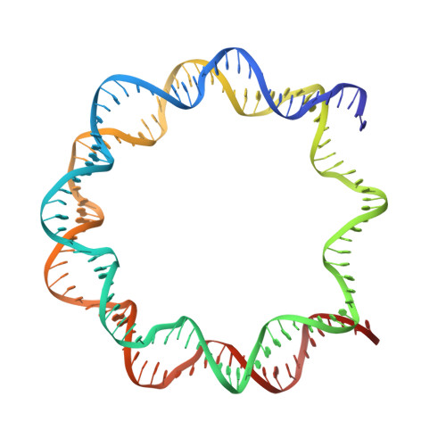
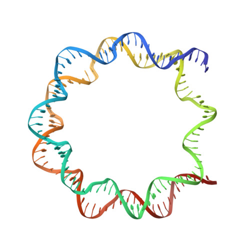
Structural insight into the sequence dependence of nucleosome positioning

Wu, B.Mohideen, K.Vasudevan, D.Davey, C.A.

(2010) Structure 18: 528-536

  • DOI: https://doi.org/10.1016/j.str.2010.01.015
  • Primary Citation Related Structures: 
    3LEL

  • PubMed Abstract: 

    Nucleosome positioning displays sequence dependency and contributes to genomic regulation in a site-specific manner. We solved the structures of nucleosome core particle composed of strong positioning TTTAA elements flanking the nucleosome center. The positioning strength of the super flexible TA dinucleotide is consistent with its observed central location within minor groove inward regions, where it can contribute maximally to energetically challenging minor groove bending, kinking and compression. The marked preference for TTTAA and positioning power of the site 1.5 double helix turns from the nucleosome center relates to a unique histone protein motif at this location, which enforces a sustained, extremely narrow minor groove via a hydrophobic "sugar clamp." Our analysis sheds light on the basis of nucleosome positioning and indicates that the histone octamer has evolved not to fully minimize sequence discrimination in DNA binding.


  • Organizational Affiliation
    • Division of Structural and Computational Biology, School of Biological Sciences, Nanyang Technological University, 60 Nanyang Drive, Singapore 637551.

Macromolecule Content 

  • Total Structure Weight: 401.12 kDa 
  • Atom Count: 24,335 
  • Modeled Residue Count: 2,128 
  • Deposited Residue Count: 2,552 
  • Unique protein chains: 4
  • Unique nucleic acid chains: 2

Macromolecules


Find similar proteins by:|  3D Structure
Entity ID: 1
MoleculeChains  Sequence LengthOrganismDetailsImage
Histone H3.2
A, E, K, O
136Xenopus laevisMutation(s): 0 
UniProt
Find proteins for P84233 (Xenopus laevis)
Explore P84233 
Go to UniProtKB:  P84233
Entity Groups
Sequence Clusters30% Identity50% Identity70% Identity90% Identity95% Identity100% Identity
UniProt GroupP84233
Sequence Annotations
Expand
Reference Sequence
Find similar proteins by:|  3D Structure
Entity ID: 2
MoleculeChains  Sequence LengthOrganismDetailsImage
Histone H4
B, F, L, P
102Xenopus laevisMutation(s): 0 
UniProt
Find proteins for P62799 (Xenopus laevis)
Explore P62799 
Go to UniProtKB:  P62799
Entity Groups
Sequence Clusters30% Identity50% Identity70% Identity90% Identity95% Identity100% Identity
UniProt GroupP62799
Sequence Annotations
Expand
Reference Sequence
Find similar proteins by:|  3D Structure
Entity ID: 3
MoleculeChains  Sequence LengthOrganismDetailsImage
Histone H2A
C, G, M, Q
128Xenopus laevisMutation(s): 0 
UniProt
Find proteins for Q6AZJ8 (Xenopus laevis)
Explore Q6AZJ8 
Go to UniProtKB:  Q6AZJ8
Entity Groups
Sequence Clusters30% Identity50% Identity70% Identity90% Identity95% Identity100% Identity
UniProt GroupQ6AZJ8
Sequence Annotations
Expand
Reference Sequence
Find similar proteins by:|  3D Structure
Entity ID: 4
MoleculeChains  Sequence LengthOrganismDetailsImage
Histone H2B 1.1
D, H, N, R
125Xenopus laevisMutation(s): 0 
UniProt
Find proteins for P02281 (Xenopus laevis)
Explore P02281 
Go to UniProtKB:  P02281
Entity Groups
Sequence Clusters30% Identity50% Identity70% Identity90% Identity95% Identity100% Identity
UniProt GroupP02281
Sequence Annotations
Expand
Reference Sequence
Find similar nucleic acids by:  (by identity cutoff) 
Entity ID: 5
MoleculeChains LengthOrganismImage
147-MER DNA
I, S
147N/A
Sequence Annotations
Expand
Reference Sequence
Find similar nucleic acids by:  (by identity cutoff) 
Entity ID: 6
MoleculeChains LengthOrganismImage
147-MER DNA
J, T
147N/A
Sequence Annotations
Expand
Reference Sequence

Small Molecules

Ligands 1 Unique
IDChains Name / Formula / InChI Key2D Diagram3D Interactions
MN

Query on MN


Download:Ideal Coordinates CCD File
AA [auth I]
AB [auth T]
BA [auth J]
BB [auth T]
CA [auth J]
AA [auth I],
AB [auth T],
BA [auth J],
BB [auth T],
CA [auth J],
DA [auth J],
EA [auth J],
FA [auth J],
GA [auth J],
HA [auth J],
IA [auth J],
JA [auth N],
KA [auth O],
LA [auth S],
MA [auth S],
NA [auth S],
OA [auth S],
PA [auth S],
QA [auth S],
RA [auth S],
SA [auth S],
TA [auth T],
U [auth G],
UA [auth T],
V [auth I],
VA [auth T],
W [auth I],
WA [auth T],
X [auth I],
XA [auth T],
Y [auth I],
YA [auth T],
Z [auth I],
ZA [auth T]
MANGANESE (II) ION
Mn
WAEMQWOKJMHJLA-UHFFFAOYSA-N

Experimental Data & Validation

Experimental Data

  • Method: X-RAY DIFFRACTION
  • Resolution: 2.95 Å
  • R-Value Free:  0.300 (Depositor), 0.297 (DCC) 
  • R-Value Work:  0.229 (Depositor), 0.224 (DCC) 
  • R-Value Observed: 0.231 (Depositor) 
Space Group: P 1 21 1
Unit Cell:
Length ( Å )Angle ( ˚ )
a = 106.747α = 90
b = 178.508β = 102.78
c = 110.412γ = 90
Software Package:
Software NamePurpose
MAR345dtbdata collection
AMoREphasing
REFMACrefinement
MOSFLMdata reduction
SCALAdata scaling

Structure Validation

View Full Validation Report


Entry History 

Deposition Data

Revision History  (Full details and data files)

  • Version 1.0: 2010-05-19
    Type: Initial release
  • Version 1.1: 2011-07-13
    Changes: Version format compliance
  • Version 1.2: 2023-11-01
    Changes: Data collection, Database references, Derived calculations, Refinement description