Mechanism of ligand-gated potassium efflux in bacterial pathogens.
Roosild, T.P., Castronovo, S., Healy, J., Miller, S., Pliotas, C., Rasmussen, T., Bartlett, W., Conway, S.J., Booth, I.R.(2010) Proc Natl Acad Sci U S A 107: 19784-19789
- PubMed: 21041667 Search on PubMedSearch on PubMed Central
- DOI: https://doi.org/10.1073/pnas.1012716107
- Primary Citation Related Structures:
3L9W, 3L9X - PubMed Abstract:
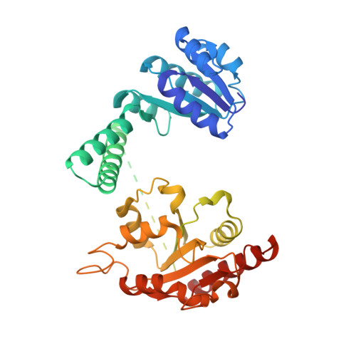
Gram negative pathogens are protected against toxic electrophilic compounds by glutathione-gated potassium efflux systems (Kef) that modulate cytoplasmic pH. We have elucidated the mechanism of gating through structural and functional analysis of Escherichia coli KefC. The revealed mechanism can explain how subtle chemical differences in glutathione derivatives can produce opposite effects on channel function. Kef channels are regulated by potassium transport and NAD-binding (KTN) domains that sense both reduced glutathione, which inhibits Kef activity, and glutathione adducts that form during electrophile detoxification and activate Kef. We find that reduced glutathione stabilizes an interdomain association between two KTN folds, whereas large adducts sterically disrupt this interaction. F441 is identified as the pivotal residue discriminating between reduced glutathione and its conjugates. We demonstrate a major structural change on the binding of an activating ligand to a KTN-domain protein. Analysis of the regulatory interactions suggests strategies to disrupt pathogen potassium and pH homeostasis.
- Drug Development Department, Nevada Cancer Institute, Las Vegas, NV 89135, USA. troosild@nvcancer.org
Organizational Affiliation: