3L6X

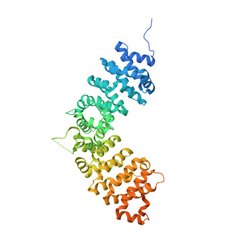
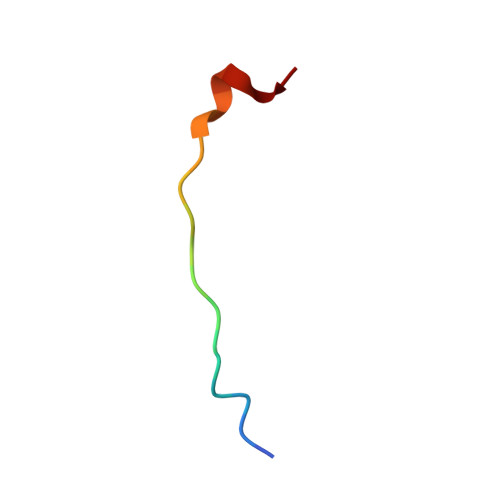
Crystal structure of p120 catenin in complex with E-cadherin


Experimental Data Snapshot

  • Method: X-RAY DIFFRACTION
  • Resolution: 2.40 Å
  • R-Value Free: 0.234 
  • R-Value Work: 0.201 
  • R-Value Observed: 0.203 

Starting Model: experimental
View more details

wwPDB Validation   3D Report Full Report


This is version 1.3 of the entry. See complete history


Literature

Dynamic and static interactions between p120 catenin and E-cadherin regulate the stability of cell-cell adhesion.

Ishiyama, N.Lee, S.H.Liu, S.Li, G.Y.Smith, M.J.Reichardt, L.F.Ikura, M.

(2010) Cell 141: 117-128

  • DOI: https://doi.org/10.1016/j.cell.2010.01.017
  • Primary Citation of Related Structures:  
    3L6X, 3L6Y

  • PubMed Abstract: 

    The association of p120 catenin (p120) with the juxtamembrane domain (JMD) of the cadherin cytoplasmic tail is critical for the surface stability of cadherin-catenin cell-cell adhesion complexes. Here, we present the crystal structure of p120 isoform 4A in complex with the JMD core region (JMD(core)) of E-cadherin. The p120 armadillo repeat domain contains modular binding pockets that are complementary to electrostatic and hydrophobic properties of the JMD(core). Single-residue mutations within the JMD(core)-binding site of p120 abolished its interaction with E- and N-cadherins in vitro and in cultured cells. These mutations of p120 enabled us to clearly differentiate between N-cadherin-dependent and -independent steps of neuronal dendritic spine morphogenesis crucial for synapse development. NMR studies revealed that p120 regulates the stability of cadherin-mediated cell-cell adhesion by associating with the majority of the JMD, including residues implicated in clathrin-mediated endocytosis and Hakai-dependent ubiquitination of E-cadherin, through its discrete "dynamic" and "static" binding sites.


  • Organizational Affiliation

    Division of Signaling Biology, Ontario Cancer Institute, ON, Canada.


Macromolecules
Find similar proteins by:  (by identity cutoff)  |  3D Structure
Entity ID: 1
MoleculeChains Sequence LengthOrganismDetailsImage
Catenin delta-1584Homo sapiensMutation(s): 0 
Gene Names: CTNND1KIAA0384
UniProt & NIH Common Fund Data Resources
Find proteins for O60716 (Homo sapiens)
Explore O60716 
Go to UniProtKB:  O60716
PHAROS:  O60716
GTEx:  ENSG00000198561 
Entity Groups  
Sequence Clusters30% Identity50% Identity70% Identity90% Identity95% Identity100% Identity
UniProt GroupO60716
Sequence Annotations
Expand
  • Reference Sequence

Find similar proteins by:  Sequence   |   3D Structure  

Entity ID: 2
MoleculeChains Sequence LengthOrganismDetailsImage
E-cadherin18Homo sapiensMutation(s): 0 
Gene Names: CDH1
UniProt & NIH Common Fund Data Resources
Find proteins for P12830 (Homo sapiens)
Explore P12830 
Go to UniProtKB:  P12830
PHAROS:  P12830
GTEx:  ENSG00000039068 
Entity Groups  
Sequence Clusters30% Identity50% Identity70% Identity90% Identity95% Identity100% Identity
UniProt GroupP12830
Sequence Annotations
Expand
  • Reference Sequence
Experimental Data & Validation

Experimental Data

  • Method: X-RAY DIFFRACTION
  • Resolution: 2.40 Å
  • R-Value Free: 0.234 
  • R-Value Work: 0.201 
  • R-Value Observed: 0.203 
  • Space Group: P 31 2 1
Unit Cell:
Length ( Å )Angle ( ˚ )
a = 92.403α = 90
b = 92.403β = 90
c = 171.89γ = 120
Software Package:
Software NamePurpose
DENZOdata reduction
SCALEPACKdata scaling
PHASERphasing
REFMACrefinement
PDB_EXTRACTdata extraction
HKL-3000data collection
HKL-2000data reduction
HKL-2000data scaling

Structure Validation

View Full Validation Report


Entry History 

Deposition Data

Revision History  (Full details and data files)

  • Version 1.0: 2010-04-21
    Type: Initial release
  • Version 1.1: 2011-07-13
    Changes: Advisory, Refinement description, Version format compliance
  • Version 1.2: 2017-11-01
    Changes: Refinement description
  • Version 1.3: 2023-09-06
    Changes: Data collection, Database references, Derived calculations, Refinement description