3L6X | pdb_00003l6x

Crystal structure of p120 catenin in complex with E-cadherin


Experimental Data Snapshot

  • Method: X-RAY DIFFRACTION
  • Resolution: 2.40 Å
  • R-Value Free: 
    0.234 (Depositor), 0.221 (DCC) 
  • R-Value Work: 
    0.201 (Depositor) 
  • R-Value Observed: 
    0.203 (Depositor) 

Starting Model: experimental
View more details

wwPDB Validation 3D Report Full Report

Validation slider image for 3L6X

This is version 1.3 of the entry. See complete history

Literature

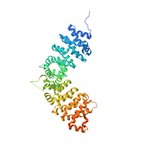
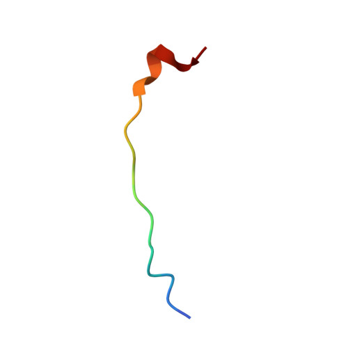
Dynamic and static interactions between p120 catenin and E-cadherin regulate the stability of cell-cell adhesion.

Ishiyama, N.Lee, S.H.Liu, S.Li, G.Y.Smith, M.J.Reichardt, L.F.Ikura, M.

(2010) Cell 141: 117-128

  • DOI: https://doi.org/10.1016/j.cell.2010.01.017
  • Primary Citation Related Structures: 
    3L6X, 3L6Y

  • PubMed Abstract: 

    The association of p120 catenin (p120) with the juxtamembrane domain (JMD) of the cadherin cytoplasmic tail is critical for the surface stability of cadherin-catenin cell-cell adhesion complexes. Here, we present the crystal structure of p120 isoform 4A in complex with the JMD core region (JMD(core)) of E-cadherin. The p120 armadillo repeat domain contains modular binding pockets that are complementary to electrostatic and hydrophobic properties of the JMD(core). Single-residue mutations within the JMD(core)-binding site of p120 abolished its interaction with E- and N-cadherins in vitro and in cultured cells. These mutations of p120 enabled us to clearly differentiate between N-cadherin-dependent and -independent steps of neuronal dendritic spine morphogenesis crucial for synapse development. NMR studies revealed that p120 regulates the stability of cadherin-mediated cell-cell adhesion by associating with the majority of the JMD, including residues implicated in clathrin-mediated endocytosis and Hakai-dependent ubiquitination of E-cadherin, through its discrete "dynamic" and "static" binding sites.


  • Organizational Affiliation
    • Division of Signaling Biology, Ontario Cancer Institute, ON, Canada.

Macromolecule Content 

  • Total Structure Weight: 67.75 kDa 
  • Atom Count: 3,664 
  • Modeled Residue Count: 452 
  • Deposited Residue Count: 602 
  • Unique protein chains: 2

Macromolecules

Find similar proteins by:|  3D Structure
Entity ID: 1
MoleculeChains  Sequence LengthOrganismDetailsImage
Catenin delta-1584Homo sapiensMutation(s): 0 
Gene Names: CTNND1KIAA0384
UniProt & NIH Common Fund Data Resources
Find proteins for O60716 (Homo sapiens)
Explore O60716 
Go to UniProtKB:  O60716
PHAROS:  O60716
GTEx:  ENSG00000198561 
Entity Groups
Sequence Clusters30% Identity50% Identity70% Identity90% Identity95% Identity100% Identity
UniProt GroupO60716
Sequence Annotations
Expand
Reference Sequence
Find similar proteins by:  Sequence   |   3D Structure  
Entity ID: 2
MoleculeChains  Sequence LengthOrganismDetailsImage
E-cadherin18Homo sapiensMutation(s): 0 
Gene Names: CDH1
UniProt & NIH Common Fund Data Resources
Find proteins for P12830 (Homo sapiens)
Explore P12830 
Go to UniProtKB:  P12830
PHAROS:  P12830
GTEx:  ENSG00000039068 
Entity Groups
Sequence Clusters30% Identity50% Identity70% Identity90% Identity95% Identity100% Identity
UniProt GroupP12830
Sequence Annotations
Expand
Reference Sequence

Experimental Data & Validation

Experimental Data

  • Method: X-RAY DIFFRACTION
  • Resolution: 2.40 Å
  • R-Value Free:  0.234 (Depositor), 0.221 (DCC) 
  • R-Value Work:  0.201 (Depositor) 
  • R-Value Observed: 0.203 (Depositor) 
Space Group: P 31 2 1
Unit Cell:
Length ( Å )Angle ( ˚ )
a = 92.403α = 90
b = 92.403β = 90
c = 171.89γ = 120
Software Package:
Software NamePurpose
DENZOdata reduction
SCALEPACKdata scaling
PHASERphasing
REFMACrefinement
PDB_EXTRACTdata extraction
HKL-3000data collection
HKL-2000data reduction
HKL-2000data scaling

Structure Validation

View Full Validation Report


Entry History 

Deposition Data

Revision History  (Full details and data files)

  • Version 1.0: 2010-04-21
    Type: Initial release
  • Version 1.1: 2011-07-13
    Changes: Advisory, Refinement description, Version format compliance
  • Version 1.2: 2017-11-01
    Changes: Refinement description
  • Version 1.3: 2023-09-06
    Changes: Data collection, Database references, Derived calculations, Refinement description