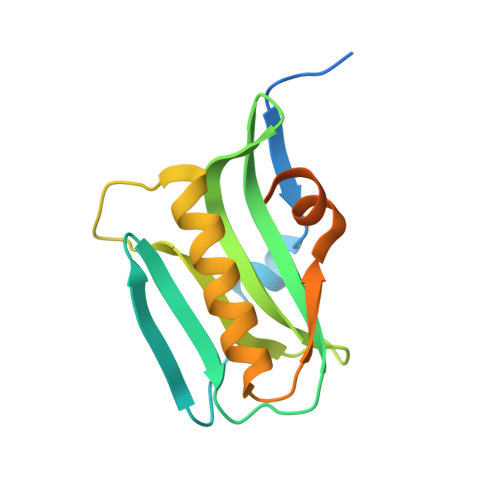
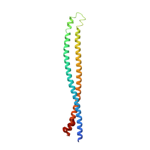
3L4Q

Structural insights into phosphoinositide 3-kinase activation by the influenza A virus NS1 protein


Experimental Data Snapshot

  • Method: X-RAY DIFFRACTION
  • Resolution: 2.30 Å
  • R-Value Free: 0.292 
  • R-Value Work: 0.232 
  • R-Value Observed: 0.235 

Starting Models: experimental
View more details

wwPDB Validation   3D Report Full Report


This is version 1.4 of the entry. See complete history


Literature

Structural insights into phosphoinositide 3-kinase activation by the influenza A virus NS1 protein

Hale, B.G.Kerry, P.S.Jackson, D.Precious, B.L.Gray, A.Killip, M.J.Randall, R.E.Russell, R.J.

(2010) Proc Natl Acad Sci U S A 107: 1954-1959

  • DOI: https://doi.org/10.1073/pnas.0910715107
  • Primary Citation of Related Structures:  
    3L4Q

  • PubMed Abstract: 

    Seasonal epidemics and periodic worldwide pandemics caused by influenza A viruses are of continuous concern. The viral nonstructural (NS1) protein is a multifunctional virulence factor that antagonizes several host innate immune defenses during infection. NS1 also directly stimulates class IA phosphoinositide 3-kinase (PI3K) signaling, an essential cell survival pathway commonly mutated in human cancers. Here, we present a 2.3-A resolution crystal structure of the NS1 effector domain in complex with the inter-SH2 (coiled-coil) domain of p85beta, a regulatory subunit of PI3K. Our data emphasize the remarkable isoform specificity of this interaction, and provide insights into the mechanism by which NS1 activates the PI3K (p85beta:p110) holoenzyme. A model of the NS1:PI3K heterotrimeric complex reveals that NS1 uses the coiled-coil as a structural tether to sterically prevent normal inhibitory contacts between the N-terminal SH2 domain of p85beta and the p110 catalytic subunit. Furthermore, in this model, NS1 makes extensive contacts with the C2/kinase domains of p110, and a small acidic alpha-helix of NS1 sits adjacent to the highly basic activation loop of the enzyme. During infection, a recombinant influenza A virus expressing NS1 with charge-disruption mutations in this acidic alpha-helix is unable to stimulate the production of phosphatidylinositol 3,4,5-trisphosphate or the phosphorylation of Akt. Despite this, the charge-disruption mutations in NS1 do not affect its ability to interact with the p85beta inter-SH2 domain in vitro. Overall, these data suggest that both direct binding of NS1 to p85beta (resulting in repositioning of the N-terminal SH2 domain) and possible NS1:p110 contacts contribute to PI3K activation.


  • Organizational Affiliation

    Biomedical Science Research Complex, University of St Andrews, North Haugh, St Andrews, Fife, KY16 9ST, United Kingdom.


Macromolecules
Find similar proteins by:  (by identity cutoff)  |  3D Structure
Entity ID: 1
MoleculeChains Sequence LengthOrganismDetailsImage
Non-structural protein 1A,
C [auth B]
164Influenza A virus (A/Puerto Rico/8/1934(H1N1))Mutation(s): 0 
Gene Names: NS
UniProt
Find proteins for P03496 (Influenza A virus (strain A/Puerto Rico/8/1934 H1N1))
Explore P03496 
Go to UniProtKB:  P03496
Entity Groups  
Sequence Clusters30% Identity50% Identity70% Identity90% Identity95% Identity100% Identity
UniProt GroupP03496
Sequence Annotations
Expand
  • Reference Sequence
Find similar proteins by:  (by identity cutoff)  |  3D Structure
Entity ID: 2
MoleculeChains Sequence LengthOrganismDetailsImage
Phosphatidylinositol 3-kinase regulatory subunit betaB [auth C],
D
170Bos taurusMutation(s): 1 
Gene Names: PIK3R2
UniProt
Find proteins for P23726 (Bos taurus)
Explore P23726 
Go to UniProtKB:  P23726
Entity Groups  
Sequence Clusters30% Identity50% Identity70% Identity90% Identity95% Identity100% Identity
UniProt GroupP23726
Sequence Annotations
Expand
  • Reference Sequence
Experimental Data & Validation

Experimental Data

  • Method: X-RAY DIFFRACTION
  • Resolution: 2.30 Å
  • R-Value Free: 0.292 
  • R-Value Work: 0.232 
  • R-Value Observed: 0.235 
  • Space Group: P 21 21 21
Unit Cell:
Length ( Å )Angle ( ˚ )
a = 57.95α = 90
b = 98.67β = 90
c = 149.94γ = 90
Software Package:
Software NamePurpose
REFMACrefinement

Structure Validation

View Full Validation Report


Entry History 

Deposition Data

Revision History  (Full details and data files)

  • Version 1.0: 2010-02-02
    Type: Initial release
  • Version 1.1: 2011-07-13
    Changes: Version format compliance
  • Version 1.2: 2014-02-12
    Changes: Database references
  • Version 1.3: 2021-11-10
    Changes: Database references, Derived calculations
  • Version 1.4: 2023-11-01
    Changes: Data collection, Refinement description