3KXP

Crystal Structure of E-2-(Acetamidomethylene)succinate Hydrolase


Experimental Data Snapshot

  • Method: X-RAY DIFFRACTION
  • Resolution: 2.26 Å
  • R-Value Free: 0.245 
  • R-Value Work: 0.204 

wwPDB Validation   3D Report Full Report


This is version 1.2 of the entry. See complete history


Literature

Structure determination and characterization of the vitamin B(6) degradative enzyme (E)-2-(acetamidomethylene)succinate hydrolase.

McCulloch, K.M.Mukherjee, T.Begley, T.P.Ealick, S.E.

(2010) Biochemistry 49: 1226-1235

  • DOI: https://doi.org/10.1021/bi901812p
  • Primary Citation of Related Structures:  
    3KXP

  • PubMed Abstract: 

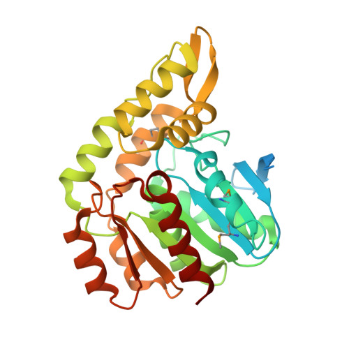
    The gene identification and kinetic characterization of (E)-2-(acetamidomethylene)succinate (E-2AMS) hydrolase has recently been described. This enzyme catalyzes the final reaction in the degradation of vitamin B(6) and produces succinic semialdehyde, acetate, ammonia, and carbon dioxide from E-2AMS. The structure of E-2AMS hydrolase was determined to 2.3 A using SAD phasing. E-2AMS hydrolase is a member of the alpha/beta hydrolase superfamily and utilizes a serine/histidine/aspartic acid catalytic triad. Mutation of either the nucleophilic serine or the aspartate resulted in inactive enzyme. Mutation of an additional serine residue in the active site causes the enzyme to be unstable and is likely structurally important. The structure also provides insight into the mechanism of hydrolysis of E-2AMS and identifies several potential catalytically important residues.


  • Organizational Affiliation

    Department of Chemistry and Chemical Biology, Cornell University, Ithaca, New York 14853, USA.


Macromolecules
Find similar proteins by:  (by identity cutoff)  |  3D Structure
Entity ID: 1
MoleculeChains Sequence LengthOrganismDetailsImage
Alpha-(N-acetylaminomethylene)succinic acid hydrolase
A, B, C, D, E
A, B, C, D, E, F, G, H, I, J, K, L
314Mesorhizobium lotiMutation(s): 0 
Gene Names: 5331
UniProt
Find proteins for Q988D4 (Mesorhizobium japonicum (strain LMG 29417 / CECT 9101 / MAFF 303099))
Explore Q988D4 
Go to UniProtKB:  Q988D4
Entity Groups  
Sequence Clusters30% Identity50% Identity70% Identity90% Identity95% Identity100% Identity
UniProt GroupQ988D4
Sequence Annotations
Expand
  • Reference Sequence
Small Molecules
Ligands 1 Unique
IDChains Name / Formula / InChI Key2D Diagram3D Interactions
CL
Query on CL

Download Ideal Coordinates CCD File 
M [auth A]
N [auth B]
O [auth C]
P [auth D]
Q [auth E]
M [auth A],
N [auth B],
O [auth C],
P [auth D],
Q [auth E],
R [auth F],
S [auth G],
T [auth H],
U [auth I],
V [auth J],
W [auth K],
X [auth L]
CHLORIDE ION
Cl
VEXZGXHMUGYJMC-UHFFFAOYSA-M
Modified Residues  1 Unique
IDChains TypeFormula2D DiagramParent
MSE
Query on MSE
A, B, C, D, E
A, B, C, D, E, F, G, H, I, J, K, L
L-PEPTIDE LINKINGC5 H11 N O2 SeMET
Experimental Data & Validation

Experimental Data

  • Method: X-RAY DIFFRACTION
  • Resolution: 2.26 Å
  • R-Value Free: 0.245 
  • R-Value Work: 0.204 
  • Space Group: P 21 21 21
Unit Cell:
Length ( Å )Angle ( ˚ )
a = 115.2α = 90
b = 178.53β = 90
c = 189.25γ = 90
Software Package:
Software NamePurpose
DENZOdata reduction
SCALEPACKdata scaling
CNSphasing
CNSrefinement
PDB_EXTRACTdata extraction

Structure Validation

View Full Validation Report


Entry History 

Deposition Data

Revision History  (Full details and data files)

  • Version 1.0: 2010-02-09
    Type: Initial release
  • Version 1.1: 2011-07-13
    Changes: Version format compliance
  • Version 1.2: 2017-11-01
    Changes: Refinement description