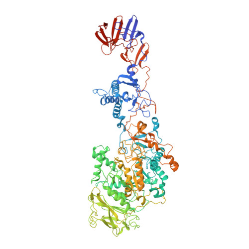
3KLK

Crystal structure of Lactobacillus reuteri N-terminally truncated glucansucrase GTF180 in triclinic apo- form


Experimental Data Snapshot

  • Method: X-RAY DIFFRACTION
  • Resolution: 1.65 Å
  • R-Value Free: 0.195 
  • R-Value Work: 0.165 
  • R-Value Observed: 0.167 

wwPDB Validation   3D Report Full Report


This is version 1.1 of the entry. See complete history


Literature

Crystal structure of a 117 kDa glucansucrase fragment provides insight into evolution and product specificity of GH70 enzymes

Vujicic-Zagar, A.Pijning, T.Kralj, S.Lopez, C.A.Eeuwema, W.Dijkhuizen, L.Dijkstra, B.W.

(2010) Proc Natl Acad Sci U S A 107: 21406-21411

  • DOI: https://doi.org/10.1073/pnas.1007531107
  • Primary Citation of Related Structures:  
    3HZ3, 3KLK, 3KLL

  • PubMed Abstract: 
  • Glucansucrases are large enzymes belonging to glycoside hydrolase family 70, which catalyze the cleavage of sucrose into fructose and glucose, with the concomitant transfer of the glucose residue to a growing α-glucan polymer. Among others, plaque-forming oral bacteria secrete these enzymes to produce α-glucans, which facilitate the adhesion of the bacteria to the tooth enamel ...

    Glucansucrases are large enzymes belonging to glycoside hydrolase family 70, which catalyze the cleavage of sucrose into fructose and glucose, with the concomitant transfer of the glucose residue to a growing α-glucan polymer. Among others, plaque-forming oral bacteria secrete these enzymes to produce α-glucans, which facilitate the adhesion of the bacteria to the tooth enamel. We determined the crystal structure of a fully active, 1,031-residue fragment encompassing the catalytic and C-terminal domains of GTF180 from Lactobacillus reuteri 180, both in the native state, and in complexes with sucrose and maltose. These structures show that the enzyme has an α-amylase-like (β/α)(8)-barrel catalytic domain that is circularly permuted compared to the catalytic domains of members of glycoside hydrolase families 13 and 77, which belong to the same GH-H superfamily. In contrast to previous suggestions, the enzyme has only one active site and one nucleophilic residue. Surprisingly, in GTF180 the peptide chain follows a "U"-path, such that four of the five domains are made up from discontiguous N- and C-terminal stretches of the peptide chain. Finally, the structures give insight into the factors that determine the different linkage types in the polymeric product.


    Organizational Affiliation

    Laboratory of Biophysical Chemistry, University of Groningen, Nijenborgh 4, 9747 AG Groningen, The Netherlands.


Macromolecules
Find similar proteins by:  (by identity cutoff)  |  3D Structure
Entity ID: 1
MoleculeChainsSequence LengthOrganismDetailsImage
Glucansucrase1,039Limosilactobacillus reuteriMutation(s): 0 
Gene Names: gtf180
EC: 2.4.1.5
UniProt
Find proteins for Q5SBN3 (Limosilactobacillus reuteri)
Explore Q5SBN3 
Go to UniProtKB:  Q5SBN3
Entity Groups  
Sequence Clusters30% Identity50% Identity70% Identity90% Identity95% Identity100% Identity
UniProt GroupQ5SBN3
Protein Feature View
Expand
  • Reference Sequence
Experimental Data & Validation

Experimental Data

  • Method: X-RAY DIFFRACTION
  • Resolution: 1.65 Å
  • R-Value Free: 0.195 
  • R-Value Work: 0.165 
  • R-Value Observed: 0.167 
  • Space Group: P 1
Unit Cell:
Length ( Å )Angle ( ˚ )
a = 58.279α = 73.3
b = 65.949β = 78.46
c = 82.506γ = 85.82
Software Package:
Software NamePurpose
SCALAdata scaling
SOLVEphasing
RESOLVEphasing
REFMACrefinement
PDB_EXTRACTdata extraction
MOSFLMdata reduction

Structure Validation

View Full Validation Report


Entry History 

Deposition Data

Revision History  (Full details and data files)

  • Version 1.0: 2010-11-03
    Type: Initial release
  • Version 1.1: 2011-07-13
    Changes: Version format compliance