3KJF

Caspase 3 Bound to a covalent inhibitor


Experimental Data Snapshot

  • Method: X-RAY DIFFRACTION
  • Resolution: 2.00 Å
  • R-Value Free: 0.206 
  • R-Value Work: 0.181 
  • R-Value Observed: 0.183 

wwPDB Validation   3D Report Full Report


Ligand Structure Quality Assessment 


This is version 1.1 of the entry. See complete history


Literature

Kinetic and structural characterization of caspase-3 and caspase-8 inhibition by a novel class of irreversible inhibitors.

Wang, Z.Watt, W.Brooks, N.A.Harris, M.S.Urban, J.Boatman, D.McMillan, M.Kahn, M.Heinrikson, R.L.Finzel, B.C.Wittwer, A.J.Blinn, J.Kamtekar, S.Tomasselli, A.G.

(2010) Biochim Biophys Acta 1804: 1817-1831

  • DOI: https://doi.org/10.1016/j.bbapap.2010.05.007
  • Primary Citation of Related Structures:  
    3KJF, 3KJN, 3KJQ

  • PubMed Abstract: 

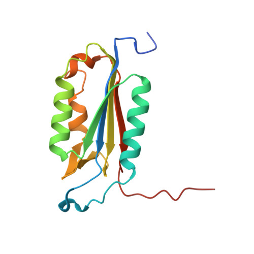
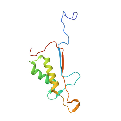
    Because of their central role in programmed cell death, the caspases are attractive targets for developing new therapeutics against cancer and autoimmunity, myocardial infarction and ischemic damage, and neurodegenerative diseases. We chose to target caspase-3, an executioner caspase, and caspase-8, an initiator caspase, based on the vast amount of information linking their functions to diseases. Through a structure-based drug design approach, a number of novel beta-strand peptidomimetic compounds were synthesized. Kinetic studies of caspase-3 and caspase-8 inhibition were carried out with these urazole ring-containing irreversible peptidomimetics and a known irreversible caspase inhibitor, Z-VAD-fmk. Using a stopped-flow fluorescence assay, we were able to determine individual kinetic parameters of caspase-3 and caspase-8 inhibition by these inhibitors. Z-VAD-fmk and the peptidomimetic inhibitors inhibit caspase-3 and caspase-8 via a three-step kinetic mechanism. Inhibition of both caspase-3 and caspase-8 by Z-VAD-fmk and of caspase-3 by the peptidomimetic inhibitors proceeds via two rapid equilibrium steps followed by a relatively fast inactivation step. However, caspase-8 inhibition by the peptidomimetics goes through a rapid equilibrium step, a slow-binding reversible step, and an extremely slow inactivation step. The crystal structures of inhibitor complexes of caspases-3 and -8 validate the design of the inhibitors by illustrating in detail how they mimic peptide substrates. One of the caspase-8 structures also shows binding at a secondary, allosteric site, providing a possible route to the development of noncovalent small molecule modulators of caspase activity.


  • Organizational Affiliation

    Oligonucleotide Therapeutics Unit, Pfizer, Inc., 620 Memorial Drive, Cambridge, MA 02139, USA. zhigang.wang@pfizer.com


Macromolecules
Find similar proteins by:  (by identity cutoff)  |  3D Structure
Entity ID: 1
MoleculeChains Sequence LengthOrganismDetailsImage
Caspase-3147Homo sapiensMutation(s): 0 
Gene Names: CASP3CPP32
EC: 3.4.22.56
UniProt & NIH Common Fund Data Resources
Find proteins for P42574 (Homo sapiens)
Explore P42574 
Go to UniProtKB:  P42574
PHAROS:  P42574
GTEx:  ENSG00000164305 
Entity Groups  
Sequence Clusters30% Identity50% Identity70% Identity90% Identity95% Identity100% Identity
UniProt GroupP42574
Sequence Annotations
Expand
  • Reference Sequence
Find similar proteins by:  (by identity cutoff)  |  3D Structure
Entity ID: 2
MoleculeChains Sequence LengthOrganismDetailsImage
Caspase-3109Homo sapiensMutation(s): 0 
Gene Names: CASP3CPP32
EC: 3.4.22.56
UniProt & NIH Common Fund Data Resources
Find proteins for P42574 (Homo sapiens)
Explore P42574 
Go to UniProtKB:  P42574
PHAROS:  P42574
GTEx:  ENSG00000164305 
Entity Groups  
Sequence Clusters30% Identity50% Identity70% Identity90% Identity95% Identity100% Identity
UniProt GroupP42574
Sequence Annotations
Expand
  • Reference Sequence
Small Molecules
Ligands 1 Unique
IDChains Name / Formula / InChI Key2D Diagram3D Interactions
B92
Query on B92

Download Ideal Coordinates CCD File 
C [auth B](3S)-3-({[(5S,10aS)-2-{(2S)-4-carboxy-2-[(phenylacetyl)amino]butyl}-1,3-dioxo-2,3,5,7,8,9,10,10a-octahydro-1H-[1,2,4]triazolo[1,2-a]cinnolin-5-yl]carbonyl}amino)-4-oxopentanoic acid
C29 H35 N5 O9
BGEXQSKGWCDSIQ-MLCQCVOFSA-N
Binding Affinity Annotations 
IDSourceBinding Affinity
B92 PDBBind:  3KJF Ki: 1.34e+4 (nM) from 1 assay(s)
Experimental Data & Validation

Experimental Data

  • Method: X-RAY DIFFRACTION
  • Resolution: 2.00 Å
  • R-Value Free: 0.206 
  • R-Value Work: 0.181 
  • R-Value Observed: 0.183 
  • Space Group: P 21 21 2
Unit Cell:
Length ( Å )Angle ( ˚ )
a = 96.69α = 90
b = 68.16β = 90
c = 44.77γ = 90
Software Package:
Software NamePurpose
HKL-2000data collection
AMoREphasing
REFMACrefinement
HKL-2000data reduction
HKL-2000data scaling

Structure Validation

View Full Validation Report


Ligand Structure Quality Assessment 


Entry History 

Deposition Data

Revision History  (Full details and data files)

  • Version 1.0: 2010-08-11
    Type: Initial release
  • Version 1.1: 2011-07-13
    Changes: Version format compliance