3KIC | pdb_00003kic

Crystal structure of adeno-associated virus serotype 3B


Experimental Data Snapshot

  • Method: X-RAY DIFFRACTION
  • Resolution: 2.60 Å
  • R-Value Free: 
    0.218 (Depositor), 0.216 (DCC) 
  • R-Value Work: 
    0.190 (Depositor), 0.191 (DCC) 
  • R-Value Observed: 
    0.190 (Depositor) 

Starting Model: experimental
View more details

wwPDB Validation 3D Report Full Report

Validation slider image for 3KIC

Ligand Structure Quality Assessment 


This is version 1.5 of the entry. See complete history

Literature

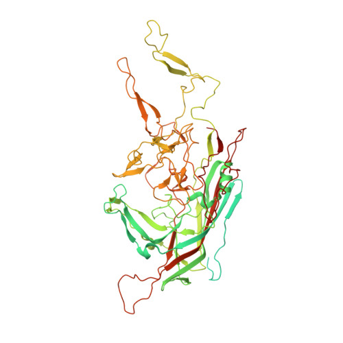
The structure of adeno-associated virus serotype 3B (AAV-3B): insights into receptor binding and immune evasion.

Lerch, T.F.Xie, Q.Chapman, M.S.

(2010) Virology 403: 26-36

  • DOI: https://doi.org/10.1016/j.virol.2010.03.027
  • Primary Citation Related Structures: 
    3KIC, 3KIE

  • PubMed Abstract: 

    Adeno-associated viruses (AAVs) are leading candidate vectors for human gene therapy. AAV serotypes have broad cellular tropism and use a variety of cellular receptors. AAV serotype 3 binds to heparan sulfate proteoglycan prior to cell entry and is serologically distinct from other serotypes. The capsid features that distinguish AAV-3B from other serotypes are poorly understood. The structure of AAV-3B has been determined to 2.6A resolution from twinned crystals of an infectious virus. The most distinctive structural features are located in regions implicated in receptor and antibody binding, providing insights into the cell entry mechanisms and antigenic nature of AAVs. We show that AAV-3B has a lower affinity for heparin than AAV-2, which can be rationalized by the distinct features of the AAV-3B capsid. The structure of AAV-3B provides an additional foundation for the future engineering of improved gene therapy vectors with modified receptor binding or antigenic characteristics.


  • Organizational Affiliation
    • Department of Biochemistry and Molecular Biology, School of Medicine, Oregon Health & Science University, Portland, OR 97239-3098, USA.

Macromolecule Content 

  • Total Structure Weight: 1,646.37 kDa 
  • Atom Count: 83,520 
  • Modeled Residue Count: 10,400 
  • Deposited Residue Count: 14,720 
  • Unique protein chains: 1

Macromolecules

Find similar proteins by:|  3D Structure
Entity ID: 1
MoleculeChains  Sequence LengthOrganismDetailsImage
Capsid protein VP1
A, B, C, D, E
A, B, C, D, E, F, G, H, I, J, K, L, M, N, O, P, Q, R, S, T
736Adeno-associated virus 3BMutation(s): 0 
Gene Names: Cap
UniProt
Find proteins for O56139 (Adeno-associated virus 3B)
Explore O56139 
Go to UniProtKB:  O56139
Entity Groups
Sequence Clusters30% Identity50% Identity70% Identity90% Identity95% Identity100% Identity
UniProt GroupO56139
Sequence Annotations
Expand
Reference Sequence

Small Molecules

Ligands 1 Unique
IDChains Name / Formula / InChI Key2D Diagram3D Interactions
D5M

Query on D5M


Download:Ideal Coordinates CCD File
AA [auth G]
BA [auth H]
CA [auth I]
DA [auth J]
EA [auth K]
AA [auth G],
BA [auth H],
CA [auth I],
DA [auth J],
EA [auth K],
FA [auth L],
GA [auth M],
HA [auth N],
IA [auth O],
JA [auth P],
KA [auth Q],
LA [auth R],
MA [auth S],
NA [auth T],
U [auth A],
V [auth B],
W [auth C],
X [auth D],
Y [auth E],
Z [auth F]
2'-DEOXYADENOSINE-5'-MONOPHOSPHATE
C10 H14 N5 O6 P
KHWCHTKSEGGWEX-RRKCRQDMSA-N

Experimental Data & Validation

Experimental Data

  • Method: X-RAY DIFFRACTION
  • Resolution: 2.60 Å
  • R-Value Free:  0.218 (Depositor), 0.216 (DCC) 
  • R-Value Work:  0.190 (Depositor), 0.191 (DCC) 
  • R-Value Observed: 0.190 (Depositor) 
Space Group: H 3
Unit Cell:
Length ( Å )Angle ( ˚ )
a = 257.704α = 90
b = 257.704β = 90
c = 603.766γ = 120
Software Package:
Software NamePurpose
DENZOdata reduction
SCALEPACKdata scaling
PHENIXrefinement
PDB_EXTRACTdata extraction
HKL-2000data collection
HKL-2000data reduction
GLRFphasing

Structure Validation

View Full Validation Report


Ligand Structure Quality Assessment 


Entry History 

Deposition Data

Revision History  (Full details and data files)

  • Version 1.0: 2010-04-28
    Type: Initial release
  • Version 1.1: 2011-07-13
    Changes: Version format compliance
  • Version 1.2: 2011-07-20
    Changes: Derived calculations, Other
  • Version 1.3: 2017-11-01
    Changes: Refinement description
  • Version 1.4: 2021-03-31
    Changes: Derived calculations, Source and taxonomy
  • Version 1.5: 2023-09-06
    Changes: Data collection, Database references, Derived calculations, Refinement description