Discovery and optimization of chromenotriazolopyrimidines as potent inhibitors of the mouse double minute 2-tumor protein 53 protein-protein interaction.
Allen, J.G., Bourbeau, M.P., Wohlhieter, G.E., Bartberger, M.D., Michelsen, K., Hungate, R., Gadwood, R.C., Gaston, R.D., Evans, B., Mann, L.W., Matison, M.E., Schneider, S., Huang, X., Yu, D., Andrews, P.S., Reichelt, A., Long, A.M., Yakowec, P., Yang, E.Y., Lee, T.A., Oliner, J.D.(2009) J Med Chem 52: 7044-7053
- PubMed: 19856920 Search on PubMed
- DOI: https://doi.org/10.1021/jm900681h
- Primary Citation Related Structures:
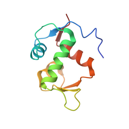
3JZK - PubMed Abstract:
Tumor protein 53 (p53) is a critical regulator of cell cycle and apoptosis that is frequently disabled in human tumors. In many tumor types, p53 is deleted or mutated, but in others p53 is inactivated by overexpression or amplification of its negative regulator mouse double minute 2 (MDM2). A high-throughput screening effort identified 6,7-bis(4-bromophenyl)-7,12-dihydro-6H-chromeno[4,3-d][1,2,4]triazolo[1,5-a]pyrimidine as a potent inhibitor of the MDM2-p53 protein-protein interaction. This screening hit was found to be chemically unstable and difficult to handle due to poor DMSO solubility. Co-crystallization with the target protein helped to direct further optimization and provided a tractable lead series of novel MDM2-p53 inhibitors. In cellular assays, these compounds were shown to upregulate p53 protein levels and p53 signaling and to cause p53-dependent inhibition of proliferation and apoptosis.
- Chemistry Research and Discovery, Amgen Inc., One Amgen Center Drive, Thousand Oaks, California 91320, USA. johallen@amgen.com
Organizational Affiliation: