3JSV

Crystal structure of mouse NEMO CoZi in complex with Lys63-linked di-ubiquitin


Experimental Data Snapshot

  • Method: X-RAY DIFFRACTION
  • Resolution: 2.70 Å
  • R-Value Free: 0.294 
  • R-Value Work: 0.250 
  • R-Value Observed: 0.250 

wwPDB Validation   3D Report Full Report


This is version 1.2 of the entry. See complete history


Literature

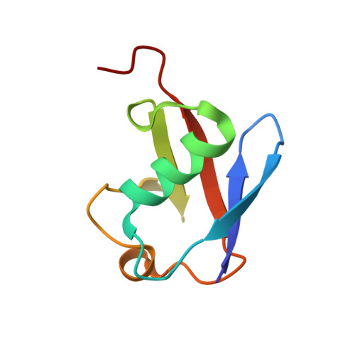
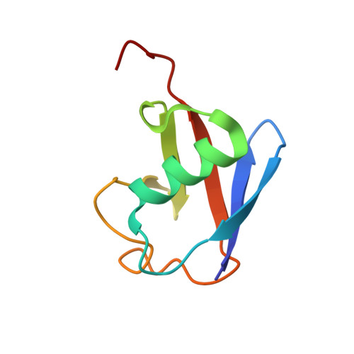
Crystal structure of the NEMO ubiquitin-binding domain in complex with Lys 63-linked di-ubiquitin

Yoshikawa, A.Sato, Y.Yamashita, M.Mimura, H.Yamagata, A.Fukai, S.

(2009) FEBS Lett 583: 3317-3322

  • DOI: https://doi.org/10.1016/j.febslet.2009.09.028
  • Primary Citation of Related Structures:  
    3JSV

  • PubMed Abstract: 

    NEMO is essential for activation of the NF-kappaB signaling pathway, which is regulated by ubiquitination of proteins. The C-terminal leucine zipper of NEMO and its adjacent coiled-coil region (CC2-LZ) reportedly bind to linear ubiquitin chains with 1 microM affinity and to Lys 63-linked chains with 100 microM affinity. Here we report the crystal structure of the CC2-LZ region of mouse NEMO in complex with Lys 63-linked di-ubiquitin (K63-Ub(2)) at 2.7A resolution. The ubiquitin-binding region consists of a 130A-long helix and forms a parallel coiled-coil dimer. The Ile 44-centered hydrophobic patch of ubiquitin is recognized in the middle of the NEMO ubiquitin-binding region. NEMO interacts with each K63-Ub(2)via a single ubiquitin-binding site, consistent with low affinity binding with K63-Ub(2).


  • Organizational Affiliation

    Structural Biology Laboratory, Life Science Division, Synchrotron Radiation Research Organization and Institute of Molecular and Cellular Biosciences, The University of Tokyo, Tokyo 113-0032, Japan.


Macromolecules
Find similar proteins by:  (by identity cutoff)  |  3D Structure
Entity ID: 1
MoleculeChains Sequence LengthOrganismDetailsImage
Ubiquitin76Homo sapiensMutation(s): 1 
UniProt & NIH Common Fund Data Resources
Find proteins for P0CG48 (Homo sapiens)
Explore P0CG48 
Go to UniProtKB:  P0CG48
PHAROS:  P0CG48
GTEx:  ENSG00000150991 
Entity Groups  
Sequence Clusters30% Identity50% Identity70% Identity90% Identity95% Identity100% Identity
UniProt GroupP0CG48
Sequence Annotations
Expand
  • Reference Sequence
Find similar proteins by:  (by identity cutoff)  |  3D Structure
Entity ID: 2
MoleculeChains Sequence LengthOrganismDetailsImage
Ubiquitin77Homo sapiensMutation(s): 1 
UniProt & NIH Common Fund Data Resources
Find proteins for P0CG48 (Homo sapiens)
Explore P0CG48 
Go to UniProtKB:  P0CG48
PHAROS:  P0CG48
GTEx:  ENSG00000150991 
Entity Groups  
Sequence Clusters30% Identity50% Identity70% Identity90% Identity95% Identity100% Identity
UniProt GroupP0CG48
Sequence Annotations
Expand
  • Reference Sequence
Find similar proteins by:  (by identity cutoff)  |  3D Structure
Entity ID: 3
MoleculeChains Sequence LengthOrganismDetailsImage
NF-kappa-B essential modulator
C, D
94Mus musculusMutation(s): 0 
Gene Names: IkbkgNemoNEMO(249-343)
UniProt
Find proteins for O88522 (Mus musculus)
Explore O88522 
Go to UniProtKB:  O88522
Entity Groups  
Sequence Clusters30% Identity50% Identity70% Identity90% Identity95% Identity100% Identity
UniProt GroupO88522
Sequence Annotations
Expand
  • Reference Sequence
Experimental Data & Validation

Experimental Data

  • Method: X-RAY DIFFRACTION
  • Resolution: 2.70 Å
  • R-Value Free: 0.294 
  • R-Value Work: 0.250 
  • R-Value Observed: 0.250 
  • Space Group: P 1 21 1
Unit Cell:
Length ( Å )Angle ( ˚ )
a = 55.38α = 90
b = 45.87β = 99.83
c = 83.29γ = 90
Software Package:
Software NamePurpose
HKL-2000data collection
MOLREPphasing
CNSrefinement
HKL-2000data reduction
HKL-2000data scaling

Structure Validation

View Full Validation Report


Entry History 

Deposition Data

Revision History  (Full details and data files)

  • Version 1.0: 2009-10-27
    Type: Initial release
  • Version 1.1: 2011-07-13
    Changes: Version format compliance
  • Version 1.2: 2021-11-10
    Changes: Database references, Derived calculations