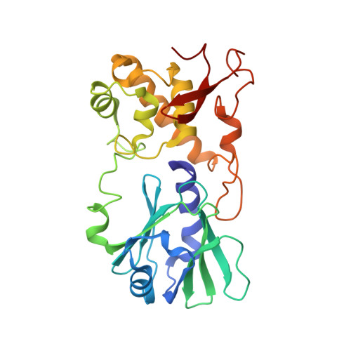
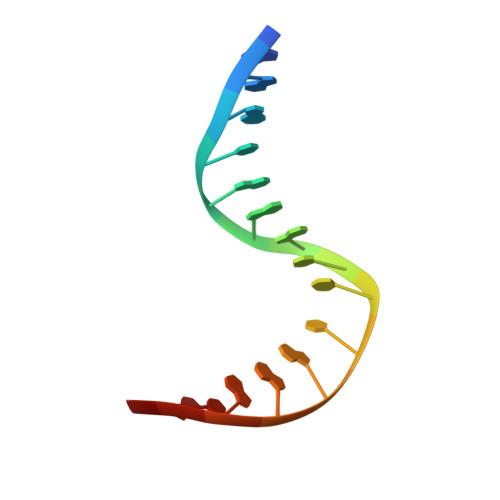
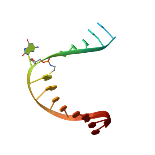
3JR5

MutM lesion recognition control complex with N174C crosslinking site


Experimental Data Snapshot

  • Method: X-RAY DIFFRACTION
  • Resolution: 1.70 Å
  • R-Value Free: 0.185 
  • R-Value Work: 0.165 
  • R-Value Observed: 0.166 

Starting Model: experimental
View more details

wwPDB Validation   3D Report Full Report


This is version 1.4 of the entry. See complete history


Literature

Entrapment and structure of an extrahelical guanine attempting to enter the active site of a bacterial DNA glycosylase, MutM.

Qi, Y.Spong, M.C.Nam, K.Karplus, M.Verdine, G.L.

(2010) J Biol Chem 285: 1468-1478

  • DOI: https://doi.org/10.1074/jbc.M109.069799
  • Primary Citation of Related Structures:  
    3JR4, 3JR5

  • PubMed Abstract: 

    MutM, a bacterial DNA glycosylase, protects genome integrity by catalyzing glycosidic bond cleavage of 8-oxoguanine (oxoG) lesions, thereby initiating base excision DNA repair. The process of searching for and locating oxoG lesions is especially challenging, because of the close structural resemblance of oxoG to its million-fold more abundant progenitor, G. Extrusion of the target nucleobase from the DNA double helix to an extrahelical position is an essential step in lesion recognition and catalysis by MutM. Although the interactions between the extruded oxoG and the active site of MutM have been well characterized, little is known in structural detail regarding the interrogation of extruded normal DNA bases by MutM. Here we report the capture and structural elucidation of a complex in which MutM is attempting to present an undamaged G to its active site. The structure of this MutM-extrahelical G complex provides insights into the mechanism MutM employs to discriminate against extrahelical normal DNA bases and into the base extrusion process in general.


  • Organizational Affiliation

    Graduate Program in Biophysics, Harvard Medical School, Boston, Massachusetts 02115, USA.


Macromolecules

Find similar proteins by:  (by identity cutoff)  |  3D Structure
Entity ID: 1
MoleculeChains Sequence LengthOrganismDetailsImage
DNA glycosylase273Geobacillus stearothermophilusMutation(s): 1 
EC: 4.2.99.18
UniProt
Find proteins for P84131 (Geobacillus stearothermophilus)
Explore P84131 
Go to UniProtKB:  P84131
Entity Groups  
Sequence Clusters30% Identity50% Identity70% Identity90% Identity95% Identity100% Identity
UniProt GroupP84131
Sequence Annotations
Expand
  • Reference Sequence

Find similar nucleic acids by:  Sequence   |   3D Structure  

Entity ID: 2
MoleculeChains LengthOrganismImage
DNA (5'-D(*AP*GP*GP*TP*AP*GP*AP*CP*TP*CP*GP*GP*AP*CP*GP*C)-3')16N/A
Sequence Annotations
Expand
  • Reference Sequence

Find similar nucleic acids by:  Sequence   |   3D Structure  

Entity ID: 3
MoleculeChains LengthOrganismImage
DNA (5'-D(*TP*GP*CP*GP*TP*CP*CP*(OGX)P*AP*GP*TP*CP*TP*AP*CP*C)-3')16N/A
Sequence Annotations
Expand
  • Reference Sequence
Experimental Data & Validation

Experimental Data

  • Method: X-RAY DIFFRACTION
  • Resolution: 1.70 Å
  • R-Value Free: 0.185 
  • R-Value Work: 0.165 
  • R-Value Observed: 0.166 
  • Space Group: P 21 21 21
Unit Cell:
Length ( Å )Angle ( ˚ )
a = 45.343α = 90
b = 95.377β = 90
c = 102.482γ = 90
Software Package:
Software NamePurpose
DENZOdata reduction
SCALEPACKdata scaling
PHENIXrefinement
PDB_EXTRACTdata extraction
ADSCdata collection
CNSphasing

Structure Validation

View Full Validation Report


Entry History 

Deposition Data

Revision History  (Full details and data files)

  • Version 1.0: 2009-11-03
    Type: Initial release
  • Version 1.1: 2011-07-13
    Changes: Non-polymer description, Version format compliance
  • Version 1.2: 2017-11-01
    Changes: Refinement description
  • Version 1.3: 2021-10-13
    Changes: Database references, Derived calculations
  • Version 1.4: 2023-09-06
    Changes: Data collection, Refinement description