3IZZ

Models for ribosome components that are nearest neighbors to the bovine mitochondrial initiation factor2 bound to the E. Coli ribosome


Experimental Data Snapshot

  • Method: ELECTRON MICROSCOPY
  • Resolution: 10.8 Å
  • Aggregation State: PARTICLE 
  • Reconstruction Method: SINGLE PARTICLE 

wwPDB Validation   3D Report Full Report


This is version 1.3 of the entry. See complete history


Literature

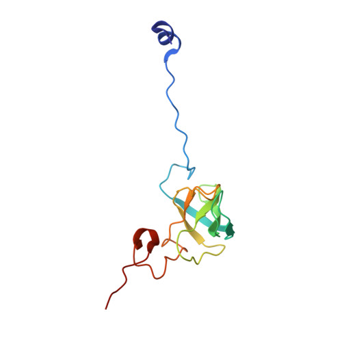
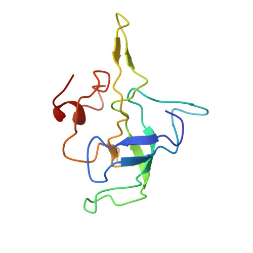
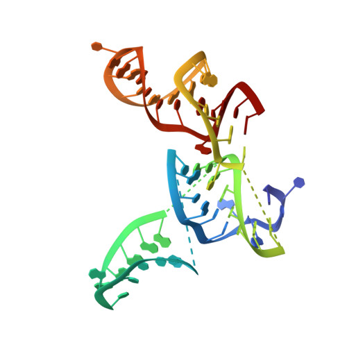
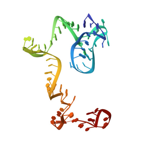
Insertion domain within mammalian mitochondrial translation initiation factor 2 serves the role of eubacterial initiation factor 1.

Yassin, A.S.Haque, M.E.Datta, P.P.Elmore, K.Banavali, N.K.Spremulli, L.L.Agrawal, R.K.

(2011) Proc Natl Acad Sci U S A 108: 3918-3923

  • DOI: https://doi.org/10.1073/pnas.1017425108
  • Primary Citation of Related Structures:  
    3IZY, 3IZZ

  • PubMed Abstract: 

    Mitochondria have their own translational machineries for the synthesis of thirteen polypeptide chains that are components of the complexes that participate in the process of oxidative phosphorylation (or ATP generation). Translation initiation in mammalian mitochondria requires two initiation factors, IF2(mt) and IF3(mt), instead of the three that are present in eubacteria. The mammalian IF2(mt) possesses a unique 37 amino acid insertion domain, which is known to be important for the formation of the translation initiation complex. We have obtained a three-dimensional cryoelectron microscopic map of the mammalian IF2(mt) in complex with initiator fMet-tRNA(iMet) and the eubacterial ribosome. We find that the 37 amino acid insertion domain interacts with the same binding site on the ribosome that would be occupied by the eubacterial initiation factor IF1, which is absent in mitochondria. Our finding suggests that the insertion domain of IF2(mt) mimics the function of eubacterial IF1, by blocking the ribosomal aminoacyl-tRNA binding site (A site) at the initiation step.


  • Organizational Affiliation

    Wadsworth Center, New York State Department of Health, Empire State Plaza, Albany, NY 12201-0509, USA.


Macromolecules

Find similar proteins by:  (by identity cutoff)  |  3D Structure
Entity ID: 4
MoleculeChains Sequence LengthOrganismDetailsImage
Protein S12 (Small Subunit)D [auth F]124Escherichia coliMutation(s): 0 
Entity Groups  
Sequence Clusters30% Identity50% Identity70% Identity90% Identity95% Identity100% Identity
Sequence Annotations
Expand
  • Reference Sequence
Find similar proteins by:  (by identity cutoff)  |  3D Structure
Entity ID: 6
MoleculeChains Sequence LengthOrganismDetailsImage
Protein L14 (Large Subunit)F [auth G]121Escherichia coliMutation(s): 0 
UniProt
Find proteins for P0ADY3 (Escherichia coli (strain K12))
Explore P0ADY3 
Go to UniProtKB:  P0ADY3
Entity Groups  
Sequence Clusters30% Identity50% Identity70% Identity90% Identity95% Identity100% Identity
UniProt GroupP0ADY3
Sequence Annotations
Expand
  • Reference Sequence
Find similar nucleic acids by:  (by identity cutoff)  |  3D Structure
Entity ID: 1
MoleculeChains LengthOrganismImage
Helix 5, 14, 15 (Small Subunit)59Escherichia coli
Sequence Annotations
Expand
  • Reference Sequence
Find similar nucleic acids by:  (by identity cutoff)  |  3D Structure
Entity ID: 2
MoleculeChains LengthOrganismImage
Helix 18 (Small Subunit)B [auth D]60Escherichia coli
Sequence Annotations
Expand
  • Reference Sequence
Find similar nucleic acids by:  (by identity cutoff)  |  3D Structure
Entity ID: 3
MoleculeChains LengthOrganismImage
Helix 44 (Small Subunit)C [auth E]100Escherichia coli
Sequence Annotations
Expand
  • Reference Sequence
Find similar nucleic acids by:  (by identity cutoff)  |  3D Structure
Entity ID: 5
MoleculeChains LengthOrganismImage
Helix 69, 71, 89, 92, 95 (Large Subunit)E [auth B]118Escherichia coli
Sequence Annotations
Expand
  • Reference Sequence
Experimental Data & Validation

Experimental Data

  • Method: ELECTRON MICROSCOPY
  • Resolution: 10.8 Å
  • Aggregation State: PARTICLE 
  • Reconstruction Method: SINGLE PARTICLE 
EM Software:
TaskSoftware PackageVersion
RECONSTRUCTIONSPIDER

Structure Validation

View Full Validation Report


Entry History 

Deposition Data

Revision History  (Full details and data files)

  • Version 1.0: 2011-03-30
    Type: Initial release
  • Version 1.1: 2011-07-13
    Changes: Version format compliance
  • Version 1.2: 2018-07-18
    Changes: Data collection
  • Version 1.3: 2024-02-21
    Changes: Data collection, Database references