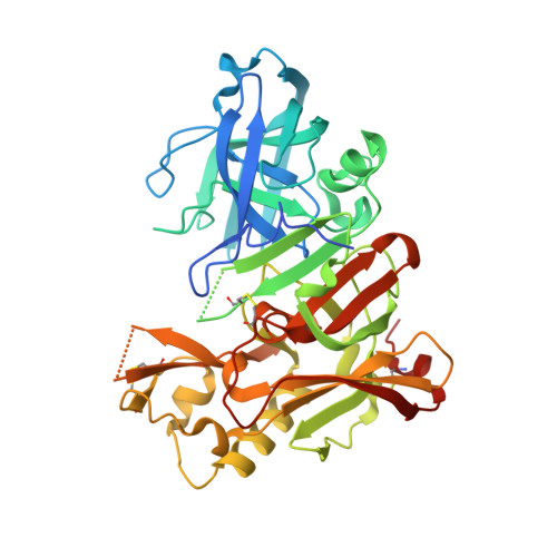
3HW1

Structure of Bace (beta secretase) in complex with ligand EV2


Experimental Data Snapshot

  • Method: X-RAY DIFFRACTION
  • Resolution: 2.48 Å
  • R-Value Free: 0.281 
  • R-Value Work: 0.226 
  • R-Value Observed: 0.226 

wwPDB Validation   3D Report Full Report


Ligand Structure Quality Assessment 


This is version 1.3 of the entry. See complete history


Literature

Fragment-Based Discovery of BACE1 Inhibitors Using Functional Assays

Godemann, R.Madden, J.Kramer, J.Smith, M.A.Fritz, U.Hesterkamp, T.Barker, J.Hoeppner, S.Hallett, D.Cesura, A.Ebneth, A.Kemp, J.

(2009) Biochemistry 48: 10743-10751

  • DOI: https://doi.org/10.1021/bi901061a
  • Primary Citation of Related Structures:  
    3HVG, 3HW1

  • PubMed Abstract: 

    Novel nonpeptidic inhibitors of beta-secretase (BACE1) have been discovered by employing a fragment-based biochemical screening approach. A diverse library of 20000 low-molecular weight compounds were screened and yielded 26 novel hits that were confirmed by biochemical and surface plasmon resonance secondary assays. We describe here fragment inhibitors cocrystallized with BACE1 in a flap open and flap closed conformation as determined by X-ray crystallography.


  • Organizational Affiliation

    Evotec AG, Schnackenburgallee 114, 22525 Hamburg, Germany. robert.godemann@evotec.com


Macromolecules
Find similar proteins by:  (by identity cutoff)  |  3D Structure
Entity ID: 1
MoleculeChains Sequence LengthOrganismDetailsImage
Beta-secretase 1
A, B, C
411Homo sapiensMutation(s): 2 
Gene Names: BACE1
EC: 3.4.23.46
UniProt & NIH Common Fund Data Resources
Find proteins for P56817 (Homo sapiens)
Explore P56817 
Go to UniProtKB:  P56817
PHAROS:  P56817
GTEx:  ENSG00000186318 
Entity Groups  
Sequence Clusters30% Identity50% Identity70% Identity90% Identity95% Identity100% Identity
UniProt GroupP56817
Sequence Annotations
Expand
  • Reference Sequence
Small Molecules
Binding Affinity Annotations 
IDSourceBinding Affinity
EV2 PDBBind:  3HW1 IC50: 8.70e+5 (nM) from 1 assay(s)
Binding MOAD:  3HW1 IC50: 8.70e+5 (nM) from 1 assay(s)
Experimental Data & Validation

Experimental Data

  • Method: X-RAY DIFFRACTION
  • Resolution: 2.48 Å
  • R-Value Free: 0.281 
  • R-Value Work: 0.226 
  • R-Value Observed: 0.226 
  • Space Group: C 1 2 1
Unit Cell:
Length ( Å )Angle ( ˚ )
a = 230.625α = 90
b = 99.88β = 103.12
c = 62.9γ = 90
Software Package:
Software NamePurpose
SLSPXdata collection
REFMACrefinement
XDSdata reduction
XSCALEdata scaling

Structure Validation

View Full Validation Report


Ligand Structure Quality Assessment 


Entry History 

Deposition Data

Revision History  (Full details and data files)

  • Version 1.0: 2009-11-03
    Type: Initial release
  • Version 1.1: 2011-07-13
    Changes: Advisory, Refinement description, Version format compliance
  • Version 1.2: 2021-11-10
    Changes: Database references, Derived calculations
  • Version 1.3: 2023-11-01
    Changes: Data collection, Refinement description