3HW1 | pdb_00003hw1

Structure of Bace (beta secretase) in complex with ligand EV2


Experimental Data Snapshot

  • Method: X-RAY DIFFRACTION
  • Resolution: 2.48 Å
  • R-Value Free: 
    0.281 (Depositor), 0.279 (DCC) 
  • R-Value Work: 
    0.226 (Depositor), 0.226 (DCC) 
  • R-Value Observed: 
    0.226 (Depositor) 

Starting Model: experimental
View more details

wwPDB Validation 3D Report Full Report

Validation slider image for 3HW1

Ligand Structure Quality Assessment 


This is version 1.4 of the entry. See complete history

Literature

Fragment-Based Discovery of BACE1 Inhibitors Using Functional Assays

Godemann, R.Madden, J.Kramer, J.Smith, M.A.Fritz, U.Hesterkamp, T.Barker, J.Hoeppner, S.Hallett, D.Cesura, A.Ebneth, A.Kemp, J.

(2009) Biochemistry 48: 10743-10751

  • DOI: https://doi.org/10.1021/bi901061a
  • Primary Citation Related Structures: 
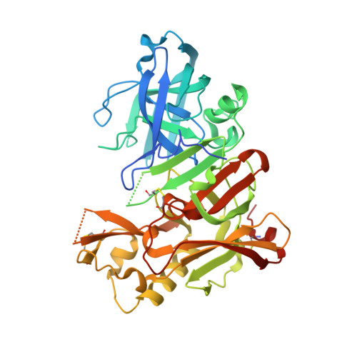
    3HVG, 3HW1

  • PubMed Abstract: 

    Novel nonpeptidic inhibitors of beta-secretase (BACE1) have been discovered by employing a fragment-based biochemical screening approach. A diverse library of 20000 low-molecular weight compounds were screened and yielded 26 novel hits that were confirmed by biochemical and surface plasmon resonance secondary assays. We describe here fragment inhibitors cocrystallized with BACE1 in a flap open and flap closed conformation as determined by X-ray crystallography.


  • Organizational Affiliation
    • Evotec AG, Schnackenburgallee 114, 22525 Hamburg, Germany. robert.godemann@evotec.com

Macromolecule Content 

  • Total Structure Weight: 138.12 kDa 
  • Atom Count: 8,985 
  • Modeled Residue Count: 1,116 
  • Deposited Residue Count: 1,233 
  • Unique protein chains: 1

Macromolecules

Find similar proteins by:|  3D Structure
Entity ID: 1
MoleculeChains  Sequence LengthOrganismDetailsImage
Beta-secretase 1
A, B, C
411Homo sapiensMutation(s): 2 
Gene Names: BACE1
EC: 3.4.23.46
UniProt & NIH Common Fund Data Resources
Find proteins for P56817 (Homo sapiens)
Explore P56817 
Go to UniProtKB:  P56817
PHAROS:  P56817
GTEx:  ENSG00000186318 
Entity Groups
Sequence Clusters30% Identity50% Identity70% Identity90% Identity95% Identity100% Identity
UniProt GroupP56817
Sequence Annotations
Expand
Reference Sequence

Experimental Data & Validation

Experimental Data

  • Method: X-RAY DIFFRACTION
  • Resolution: 2.48 Å
  • R-Value Free:  0.281 (Depositor), 0.279 (DCC) 
  • R-Value Work:  0.226 (Depositor), 0.226 (DCC) 
  • R-Value Observed: 0.226 (Depositor) 
Space Group: C 1 2 1
Unit Cell:
Length ( Å )Angle ( ˚ )
a = 230.625α = 90
b = 99.88β = 103.12
c = 62.9γ = 90
Software Package:
Software NamePurpose
SLSPXdata collection
REFMACrefinement
XDSdata reduction
XSCALEdata scaling

Structure Validation

View Full Validation Report


Ligand Structure Quality Assessment 


Entry History 

Deposition Data

Revision History  (Full details and data files)

  • Version 1.0: 2009-11-03
    Type: Initial release
  • Version 1.1: 2011-07-13
    Changes: Advisory, Refinement description, Version format compliance
  • Version 1.2: 2021-11-10
    Changes: Database references, Derived calculations
  • Version 1.3: 2023-11-01
    Changes: Data collection, Refinement description
  • Version 1.4: 2024-10-30
    Changes: Structure summary