3HVE | pdb_00003hve

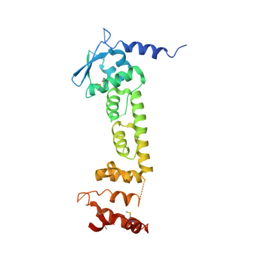
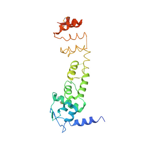
Structures of SPOP-Substrate Complexes: Insights into Molecular Architectures of BTB-Cul3 Ubiquitin Ligases: GigaxoninBTB/3-box


Experimental Data Snapshot

  • Method: X-RAY DIFFRACTION
  • Resolution: 2.80 Å
  • R-Value Free: 
    0.333 (Depositor), 0.328 (DCC) 
  • R-Value Work: 
    0.302 (Depositor), 0.297 (DCC) 

wwPDB Validation 3D Report Full Report

Validation slider image for 3HVE

This is version 1.3 of the entry. See complete history. 

Literature

Structures of SPOP-substrate complexes: insights into molecular architectures of BTB-Cul3 ubiquitin ligases.

Zhuang, M., Calabrese, M.F., Liu, J., Waddell, M.B., Nourse, A., Hammel, M., Miller, D.J., Walden, H., Duda, D.M., Seyedin, S.N., Hoggard, T., Harper, J.W., White, K.P., Schulman, B.A.

(2009) Mol Cell 36: 39-50

  • DOI: https://doi.org/10.1016/j.molcel.2009.09.022
  • Primary Citation Related Structures: 
    3HQH, 3HQI, 3HQL, 3HQM, 3HSV, 3HTM, 3HU6, 3HVE, 3IVQ, 3IVV

  • PubMed Abstract: 

    In the largest E3 ligase subfamily, Cul3 binds a BTB domain, and an associated protein-interaction domain such as MATH recruits substrates for ubiquitination. Here, we present biochemical and structural analyses of the MATH-BTB protein, SPOP. We define a SPOP-binding consensus (SBC) and determine structures revealing recognition of SBCs from the phosphatase Puc, the transcriptional regulator Ci, and the chromatin component MacroH2A. We identify a dimeric SPOP-Cul3 assembly involving a conserved helical structure C-terminal of BTB domains, which we call "3-box" due to its facilitating Cul3 binding and its resemblance to F-/SOCS-boxes in other cullin-based E3s. Structural flexibility between the substrate-binding MATH and Cul3-binding BTB/3-box domains potentially allows a SPOP dimer to engage multiple SBCs found within a single substrate, such as Puc. These studies provide a molecular understanding of how MATH-BTB proteins recruit substrates to Cul3 and how their dimerization and conformational variability may facilitate avid interactions with diverse substrates.


  • Organizational Affiliation: 
    • Department of Structural Biology, St. Jude Children's Research Hospital, Memphis, TN 38105, USA.

Macromolecule Content 

  • Total Structure Weight: 57.35 kDa 
  • Atom Count: 3,049 
  • Modeled Residue Count: 411 
  • Deposited Residue Count: 512 
  • Unique protein chains: 2

Macromolecules

Find similar proteins by:|  3D Structure
Entity ID: 1
MoleculeChains  Sequence LengthOrganismDetailsImage
Gigaxonin256Homo sapiensMutation(s): 0 
Gene Names: GAN, GAN1, KLHL16
UniProt & NIH Common Fund Data Resources
Find proteins for Q9H2C0 (Homo sapiens)
Explore Q9H2C0 
Go to UniProtKB:  Q9H2C0
PHAROS:  Q9H2C0
GTEx:  ENSG00000261609 
Entity Groups
Sequence Clusters30% Identity50% Identity70% Identity90% Identity95% Identity100% Identity
UniProt GroupQ9H2C0
Sequence Annotations
Expand
Reference Sequence
Find similar proteins by:|  3D Structure
Entity ID: 2
MoleculeChains  Sequence LengthOrganismDetailsImage
Gigaxonin256Homo sapiensMutation(s): 0 
Gene Names: GAN, GAN1, KLHL16
UniProt & NIH Common Fund Data Resources
Find proteins for Q9H2C0 (Homo sapiens)
Explore Q9H2C0 
Go to UniProtKB:  Q9H2C0
PHAROS:  Q9H2C0
GTEx:  ENSG00000261609 
Entity Groups
Sequence Clusters30% Identity50% Identity70% Identity90% Identity95% Identity100% Identity
UniProt GroupQ9H2C0
Sequence Annotations
Expand
Reference Sequence

Small Molecules

Modified Residues  2 Unique
IDChains TypeFormula2D DiagramParent
MSE
Query on MSE
A
L-PEPTIDE LINKINGC5 H11 N O2 SeMET
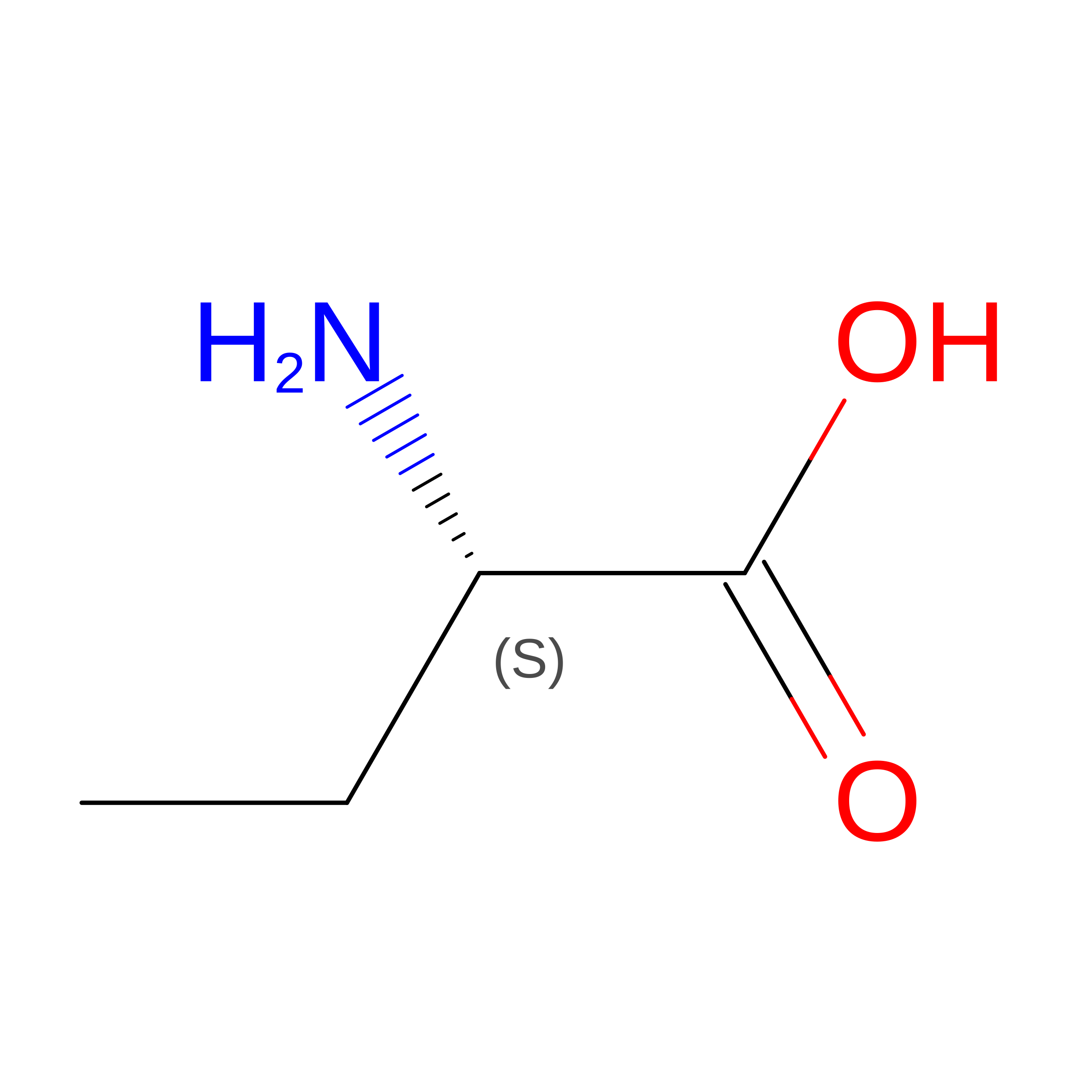
UNK
Query on UNK
B
L-PEPTIDE LINKINGC4 H9 N O2

--

Experimental Data & Validation

Experimental Data

  • Method: X-RAY DIFFRACTION
  • Resolution: 2.80 Å
  • R-Value Free:  0.333 (Depositor), 0.328 (DCC) 
  • R-Value Work:  0.302 (Depositor), 0.297 (DCC) 
Space Group: P 1 21 1
Unit Cell:
Length ( Å )Angle ( ˚ )
a = 46.48α = 90
b = 55.6β = 91.1
c = 120.6γ = 90
Software Package:
Software NamePurpose
SOLVEphasing
CNSrefinement
HKL-2000data reduction
HKL-2000data scaling

Structure Validation

View Full Validation Report


Entry History 

Deposition Data

Revision History  (Full details and data files)

  • Version 1.0: 2009-10-27
    Type: Initial release
  • Version 1.1: 2011-07-13
    Changes: Version format compliance
  • Version 1.2: 2018-06-27
    Changes: Data collection
  • Version 1.3: 2024-10-30
    Changes: Data collection, Database references, Derived calculations, Structure summary