3HOS

Crystal structure of the mariner Mos1 paired end complex with Mg


Experimental Data Snapshot

  • Method: X-RAY DIFFRACTION
  • Resolution: 3.50 Å
  • R-Value Free: 0.279 
  • R-Value Work: 0.219 
  • R-Value Observed: 0.222 

wwPDB Validation   3D Report Full Report


This is version 1.4 of the entry. See complete history


Literature

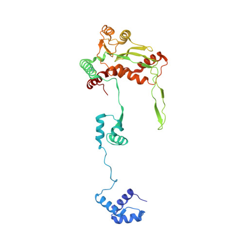
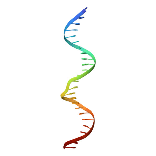
Molecular architecture of the Mos1 paired-end complex: the structural basis of DNA transposition in a eukaryote

Richardson, J.M.Colloms, S.D.Finnegan, D.J.Walkinshaw, M.D.

(2009) Cell 138: 1096-1108

  • DOI: https://doi.org/10.1016/j.cell.2009.07.012
  • Primary Citation of Related Structures:  
    3HOS, 3HOT

  • PubMed Abstract: 

    A key step in cut-and-paste DNA transposition is the pairing of transposon ends before the element is excised and inserted at a new site in its host genome. Crystallographic analyses of the paired-end complex (PEC) formed from precleaved transposon ends and the transposase of the eukaryotic element Mos1 reveals two parallel ends bound to a dimeric enzyme. The complex has a trans arrangement, with each transposon end recognized by the DNA binding region of one transposase monomer and by the active site of the other monomer. Two additional DNA duplexes in the crystal indicate likely binding sites for flanking DNA. Biochemical data provide support for a model of the target capture complex and identify Arg186 to be critical for target binding. Mixing experiments indicate that a transposase dimer initiates first-strand cleavage and suggest a pathway for PEC formation.


  • Organizational Affiliation

    School of Biological Sciences, University of Edinburgh, Edinburgh, Scotland. jrichard@staffmail.ed.ac.uk


Macromolecules

Find similar proteins by:  (by identity cutoff)  |  3D Structure
Entity ID: 1
MoleculeChains Sequence LengthOrganismDetailsImage
Transposable element mariner, complete cds
A, B
345Drosophila mauritianaMutation(s): 1 
Gene Names: T
EC: 2.7.7
UniProt
Find proteins for Q7JQ07 (Drosophila mauritiana)
Explore Q7JQ07 
Go to UniProtKB:  Q7JQ07
Entity Groups  
Sequence Clusters30% Identity50% Identity70% Identity90% Identity95% Identity100% Identity
UniProt GroupQ7JQ07
Sequence Annotations
Expand
  • Reference Sequence

Find similar nucleic acids by:  Sequence   |   3D Structure  

Entity ID: 2
MoleculeChains LengthOrganismImage
Mos1 NTS inverted repeat DNA
C, E, G
25N/A
Sequence Annotations
Expand
  • Reference Sequence
Find similar nucleic acids by:  (by identity cutoff)  |  3D Structure
Entity ID: 3
MoleculeChains LengthOrganismImage
Mos1 TS inverted repeat DNA
D, F, H
28N/A
Sequence Annotations
Expand
  • Reference Sequence
Experimental Data & Validation

Experimental Data

  • Method: X-RAY DIFFRACTION
  • Resolution: 3.50 Å
  • R-Value Free: 0.279 
  • R-Value Work: 0.219 
  • R-Value Observed: 0.222 
  • Space Group: P 1 21 1
Unit Cell:
Length ( Å )Angle ( ˚ )
a = 121.232α = 90
b = 85.033β = 98.93
c = 131.273γ = 90
Software Package:
Software NamePurpose
MOSFLMdata reduction
SCALAdata scaling
PHASERphasing
DMphasing
REFMACrefinement
PDB_EXTRACTdata extraction
ADSCdata collection

Structure Validation

View Full Validation Report


Entry History 

Deposition Data

Revision History  (Full details and data files)

  • Version 1.0: 2009-09-22
    Type: Initial release
  • Version 1.1: 2011-07-13
    Changes: Advisory, Version format compliance
  • Version 1.2: 2013-10-16
    Changes: Derived calculations
  • Version 1.3: 2021-11-10
    Changes: Database references, Derived calculations
  • Version 1.4: 2023-11-01
    Changes: Data collection, Refinement description