3HO4 | pdb_00003ho4

Crystal structure of Hedgehog-interacting protein (HHIP)


Experimental Data Snapshot

  • Method: X-RAY DIFFRACTION
  • Resolution: 3.10 Å
  • R-Value Free: 
    0.252 (Depositor), 0.245 (DCC) 
  • R-Value Work: 
    0.217 (Depositor), 0.210 (DCC) 
  • R-Value Observed: 
    0.219 (Depositor) 

wwPDB Validation 3D Report Full Report

Validation slider image for 3HO4

This is version 1.2 of the entry. See complete history

Literature

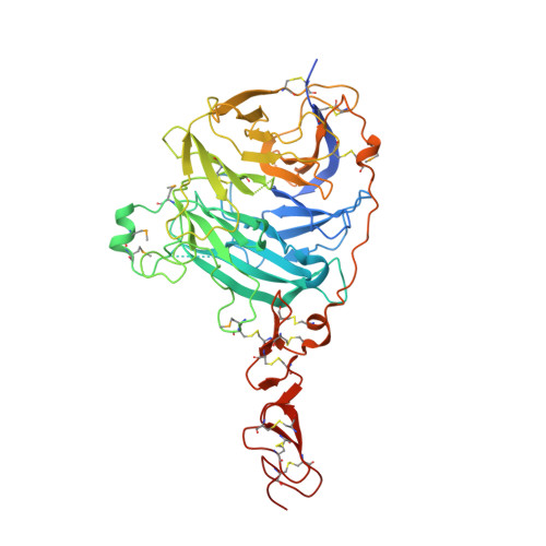
The structure of SHH in complex with HHIP reveals a recognition role for the Shh pseudo active site in signaling.

Bosanac, I.Maun, H.R.Scales, S.J.Wen, X.Lingel, A.Bazan, J.F.de Sauvage, F.J.Hymowitz, S.G.Lazarus, R.A.

(2009) Nat Struct Mol Biol 16: 691-697

  • DOI: https://doi.org/10.1038/nsmb.1632
  • Primary Citation Related Structures: 
    3HO3, 3HO4, 3HO5

  • PubMed Abstract: 

    Hedgehog (Hh) signaling is crucial for many aspects of embryonic development, whereas dysregulation of this pathway is associated with several types of cancer. Hedgehog-interacting protein (Hhip) is a surface receptor antagonist that is equipotent against all three mammalian Hh homologs. The crystal structures of human HHIP alone and bound to Sonic hedgehog (SHH) now reveal that HHIP is comprised of two EGF domains and a six-bladed beta-propeller domain. In the complex structure, a critical loop from HHIP binds the pseudo active site groove of SHH and directly coordinates its Zn2+ cation. Notably, sequence comparisons of this SHH binding loop with the Hh receptor Patched (Ptc1) ectodomains and HHIP- and PTC1-peptide binding studies suggest a 'patch for Patched' at the Shh pseudo active site; thus, we propose a role for Hhip as a structural decoy receptor for vertebrate Hh.


  • Organizational Affiliation
    • Department of Structural Biology, Genentech, Inc., South San Francisco, California, USA.

Macromolecule Content 

  • Total Structure Weight: 108.83 kDa 
  • Atom Count: 6,868 
  • Modeled Residue Count: 879 
  • Deposited Residue Count: 962 
  • Unique protein chains: 1

Macromolecules

Find similar proteins by:|  3D Structure
Entity ID: 1
MoleculeChains  Sequence LengthOrganismDetailsImage
Hedgehog-interacting protein
A, B
481Homo sapiensMutation(s): 0 
Gene Names: HHIPHHIP ORFNames: UNQ5825/PRO19644HIPUNQ5825/PRO19644
UniProt & NIH Common Fund Data Resources
Find proteins for Q96QV1 (Homo sapiens)
Explore Q96QV1 
Go to UniProtKB:  Q96QV1
PHAROS:  Q96QV1
GTEx:  ENSG00000164161 
Entity Groups
Sequence Clusters30% Identity50% Identity70% Identity90% Identity95% Identity100% Identity
UniProt GroupQ96QV1
Sequence Annotations
Expand
Reference Sequence

Small Molecules

Modified Residues  1 Unique
IDChains TypeFormula2D DiagramParent
MSE
Query on MSE
A, B
L-PEPTIDE LINKINGC5 H11 N O2 SeMET

Experimental Data & Validation

Experimental Data

  • Method: X-RAY DIFFRACTION
  • Resolution: 3.10 Å
  • R-Value Free:  0.252 (Depositor), 0.245 (DCC) 
  • R-Value Work:  0.217 (Depositor), 0.210 (DCC) 
  • R-Value Observed: 0.219 (Depositor) 
Space Group: P 31 2 1
Unit Cell:
Length ( Å )Angle ( ˚ )
a = 101.042α = 90
b = 101.042β = 90
c = 304.923γ = 120
Software Package:
Software NamePurpose
Blu-Icedata collection
SHARPphasing
REFMACrefinement
HKL-2000data reduction
HKL-2000data scaling

Structure Validation

View Full Validation Report


Entry History 

Deposition Data

Revision History  (Full details and data files)

  • Version 1.0: 2009-06-23
    Type: Initial release
  • Version 1.1: 2011-07-13
    Changes: Advisory, Version format compliance
  • Version 1.2: 2024-10-16
    Changes: Data collection, Database references, Derived calculations, Structure summary