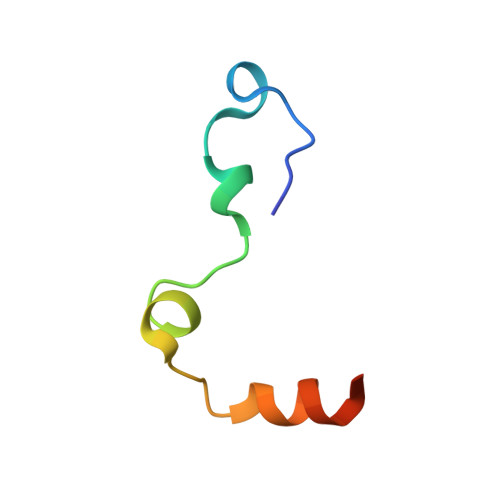
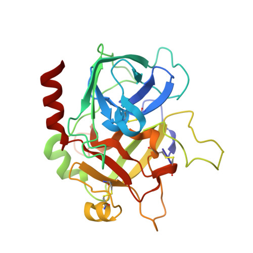
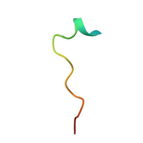
3HKI

Crystal structure of murine thrombin mutant W215A/E217A in complex with the extracellular fragment of human PAR1


Experimental Data Snapshot

  • Method: X-RAY DIFFRACTION
  • Resolution: 2.20 Å
  • R-Value Free: 0.261 
  • R-Value Work: 0.228 
  • R-Value Observed: 0.228 

Starting Model: experimental
View more details

wwPDB Validation   3D Report Full Report


Ligand Structure Quality Assessment 


This is version 1.4 of the entry. See complete history


Literature

Mechanism of the Anticoagulant Activity of Thrombin Mutant W215A/E217A.

Gandhi, P.S.Page, M.J.Chen, Z.Bush-Pelc, L.Di Cera, E.

(2009) J Biol Chem 284: 24098-24105

  • DOI: https://doi.org/10.1074/jbc.M109.025403
  • Primary Citation of Related Structures:  
    3HK3, 3HK6, 3HKI, 3HKJ

  • PubMed Abstract: 

    The thrombin mutant W215A/E217A (WE) is a potent anticoagulant both in vitro and in vivo. Previous x-ray structural studies have shown that WE assumes a partially collapsed conformation that is similar to the inactive E* form, which explains its drastically reduced activity toward substrate. Whether this collapsed conformation is genuine, rather than the result of crystal packing or the mutation introduced in the critical 215-217 beta-strand, and whether binding of thrombomodulin to exosite I can allosterically shift the E* form to the active E form to restore activity toward protein C are issues of considerable mechanistic importance to improve the design of an anticoagulant thrombin mutant for therapeutic applications. Here we present four crystal structures of WE in the human and murine forms that confirm the collapsed conformation reported previously under different experimental conditions and crystal packing. We also present structures of human and murine WE bound to exosite I with a fragment of the platelet receptor PAR1, which is unable to shift WE to the E form. These structural findings, along with kinetic and calorimetry data, indicate that WE is strongly stabilized in the E* form and explain why binding of ligands to exosite I has only a modest effect on the E*-E equilibrium for this mutant. The E* --> E transition requires the combined binding of thrombomodulin and protein C and restores activity of the mutant WE in the anticoagulant pathway.


  • Organizational Affiliation

    Department of Biochemistry and Molecular Biophysics, Washington University School of Medicine, St. Louis, Missouri 63110, USA.


Macromolecules
Find similar proteins by:  (by identity cutoff)  |  3D Structure
Entity ID: 1
MoleculeChains Sequence LengthOrganismDetailsImage
Thrombin light chain
A, D
44Mus musculusMutation(s): 0 
Gene Names: F2Cf2
EC: 3.4.21.5
UniProt & NIH Common Fund Data Resources
Find proteins for P19221 (Mus musculus)
Explore P19221 
Go to UniProtKB:  P19221
IMPC:  MGI:88380
Entity Groups  
Sequence Clusters30% Identity50% Identity70% Identity90% Identity95% Identity100% Identity
UniProt GroupP19221
Sequence Annotations
Expand
  • Reference Sequence
Find similar proteins by:  (by identity cutoff)  |  3D Structure
Entity ID: 2
MoleculeChains Sequence LengthOrganismDetailsImage
Thrombin heavy chain
B, E
258Mus musculusMutation(s): 2 
Gene Names: F2Cf2
EC: 3.4.21.5
UniProt & NIH Common Fund Data Resources
Find proteins for P19221 (Mus musculus)
Explore P19221 
Go to UniProtKB:  P19221
IMPC:  MGI:88380
Entity Groups  
Sequence Clusters30% Identity50% Identity70% Identity90% Identity95% Identity100% Identity
UniProt GroupP19221
Glycosylation
Glycosylation Sites: 1Go to GlyGen: P19221-1
Sequence Annotations
Expand
  • Reference Sequence

Find similar proteins by:  Sequence   |   3D Structure  

Entity ID: 3
MoleculeChains Sequence LengthOrganismDetailsImage
Proteinase-activated receptor 1
C, F
21Homo sapiensMutation(s): 0 
Gene Names: F2RCF2RPAR1TR
UniProt & NIH Common Fund Data Resources
Find proteins for P25116 (Homo sapiens)
Explore P25116 
Go to UniProtKB:  P25116
PHAROS:  P25116
GTEx:  ENSG00000181104 
Entity Groups  
Sequence Clusters30% Identity50% Identity70% Identity90% Identity95% Identity100% Identity
UniProt GroupP25116
Sequence Annotations
Expand
  • Reference Sequence
Experimental Data & Validation

Experimental Data

  • Method: X-RAY DIFFRACTION
  • Resolution: 2.20 Å
  • R-Value Free: 0.261 
  • R-Value Work: 0.228 
  • R-Value Observed: 0.228 
  • Space Group: P 21 21 2
Unit Cell:
Length ( Å )Angle ( ˚ )
a = 231.419α = 90
b = 50.995β = 90
c = 80.54γ = 90
Software Package:
Software NamePurpose
ADSCdata collection
MOLREPphasing
CNSrefinement
HKL-2000data reduction
SCALEPACKdata scaling

Structure Validation

View Full Validation Report


Ligand Structure Quality Assessment 


Entry History 

Deposition Data

Revision History  (Full details and data files)

  • Version 1.0: 2009-07-07
    Type: Initial release
  • Version 1.1: 2011-07-13
    Changes: Non-polymer description, Version format compliance
  • Version 1.2: 2020-07-29
    Type: Remediation
    Reason: Carbohydrate remediation
    Changes: Data collection, Derived calculations, Structure summary
  • Version 1.3: 2021-10-13
    Changes: Database references, Structure summary
  • Version 1.4: 2023-09-06
    Changes: Data collection, Refinement description