The X-ray Crystal Structure of the First RNA Recognition Motif and Site-Directed Mutagenesis Suggest a Possible HuR Redox Sensing Mechanism.
Benoit, R.M., Meisner, N.C., Kallen, J., Graff, P., Hemmig, R., Cebe, R., Ostermeier, C., Widmer, H., Auer, M.(2010) J Mol Biology 397: 1231-1244
- PubMed: 20219472 Search on PubMed
- DOI: https://doi.org/10.1016/j.jmb.2010.02.043
- Primary Citation Related Structures:
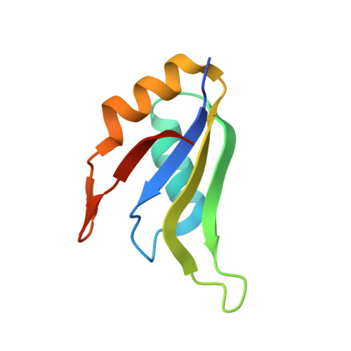
3HI9 - PubMed Abstract:
Hu-antigen R (HuR) is a ubiquitous RNA-binding protein that comprises three RNA recognition motifs (RRMs). The first two tandem RRMs are known to bind to AU-rich elements (AREs) in the 3'-untranslated region of many mRNAs. The third RRM is connected to the second RRM through a basic hinge region that contains a localization signal termed HuR nucleocytoplasmic shuttling. Binding of HuR to the ARE in the 3'-untranslated region of mRNA leads to nuclear export, stabilization, and/or translational de-repression of the mRNA, resulting in upregulation of the encoded protein. Among the various ARE binding proteins known to date, HuR is still the only known ubiquitous antagonist of posttranscriptional gene silencing by AREs. Given the wide repertoire of known and suspected targets of HuR, it is considered to be a central node in the ARE pathway. Here, the x-ray crystal structure of the first RRM of HuR (amino acids 18-99) at 2.0 A resolution is presented. The overall fold consists of two alpha-helices and a four-stranded beta-sheet, with a beta1-alpha1-beta2-beta3-alpha2-beta4 topology and a beta-hairpin between alpha2 and beta4. The asymmetric unit consists of four chains. The large crystal contact interfaces observed between chains A/B and C/D contain hydrophobic residues located at the alpha-helix side of the fold, opposite to the RNA-binding interface. This hydrophobic region structurally resembles the protein-protein interaction site of RRM domains of other proteins. Because the nature of the assumed HuR homodimerization is mechanistically not well understood to date, we used site-directed mutagenesis, analytical size-exclusion chromatography and multiangle light scattering to investigate HuR interactions via the RRM hydrophobic region. Our data indicate that in vitro, HuR RRM1 and RRM1,2 homodimerization involves a disulfide bond at cysteine 13. This homodimerization mode may have a functional significance in redox modulation of HuR activity in response to oxidative stress. Because HuR is involved in many diseases (e.g., cancer, cachexia, and inflammatory bowel disease), the presented structure may provide a basis for rational drug design.
- Novartis Institutes for BioMedical Research, Novartis Campus Forum 1, 4056 Basel, Switzerland.
Organizational Affiliation: