Crystal Structures of TbCatB and rhodesain, potential chemotherapeutic targets and major cysteine proteases of Trypanosoma brucei
Kerr, I.D., Wu, P., Marion-Tsukamaki, R., Mackey, Z.B., Brinen, L.S.(2010) PLoS Negl Trop Dis 4: e701-e701
- PubMed: 20544024 Search on PubMedSearch on PubMed Central
- DOI: https://doi.org/10.1371/journal.pntd.0000701
- Primary Citation Related Structures:
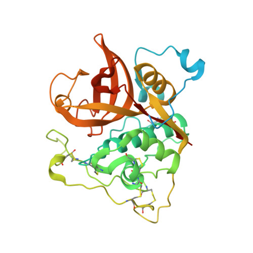
3HHI - PubMed Abstract:
Trypanosoma brucei is the etiological agent of Human African Trypanosomiasis, an endemic parasitic disease of sub-Saharan Africa. TbCatB and rhodesain are the sole Clan CA papain-like cysteine proteases produced by the parasite during infection of the mammalian host and are implicated in the progression of disease. Of considerable interest is the exploration of these two enzymes as targets for cysteine protease inhibitors that are effective against T. brucei. We have determined, by X-ray crystallography, the first reported structure of TbCatB in complex with the cathepsin B selective inhibitor CA074. In addition we report the structure of rhodesain in complex with the vinyl-sulfone K11002. The mature domain of our TbCat*CA074 structure contains unique features for a cathepsin B-like enzyme including an elongated N-terminus extending 16 residues past the predicted maturation cleavage site. N-terminal Edman sequencing reveals an even longer extension than is observed amongst the ordered portions of the crystal structure. The TbCat*CA074 structure confirms that the occluding loop, which is an essential part of the substrate-binding site, creates a larger prime side pocket in the active site cleft than is found in mammalian cathepsin B-small molecule structures. Our data further highlight enhanced flexibility in the occluding loop main chain and structural deviations from mammalian cathepsin B enzymes that may affect activity and inhibitor design. Comparisons with the rhodesain*K11002 structure highlight key differences that may impact the design of cysteine protease inhibitors as anti-trypanosomal drugs.
- Department of Cellular and Molecular Pharmacology, University of California San Francisco, San Francisco, California, United States of America.
Organizational Affiliation: