3HD3 | pdb_00003hd3

High resolution crystal structure of cruzain bound to the vinyl sulfone inhibitor SMDC-256047


Experimental Data Snapshot

  • Method: X-RAY DIFFRACTION
  • Resolution: 1.75 Å
  • R-Value Free: 
    0.176 (Depositor), 0.176 (DCC) 
  • R-Value Work: 
    0.143 (Depositor), 0.145 (DCC) 
  • R-Value Observed: 
    0.145 (Depositor) 

wwPDB Validation 3D Report Full Report

Validation slider image for 3HD3

Ligand Structure Quality Assessment 


This is version 1.3 of the entry. See complete history

Literature

Novel non-peptidic vinylsulfones targeting the S2 and S3 subsites of parasite cysteine proteases.

Bryant, C.Kerr, I.D.Debnath, M.Ang, K.K.Ratnam, J.Ferreira, R.S.Jaishankar, P.Zhao, D.Arkin, M.R.McKerrow, J.H.Brinen, L.S.Renslo, A.R.

(2009) Bioorg Med Chem Lett 19: 6218-6221

  • DOI: https://doi.org/10.1016/j.bmcl.2009.08.098
  • Primary Citation Related Structures: 
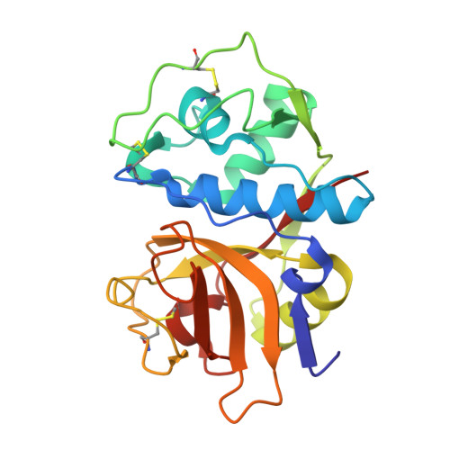
    3HD3

  • PubMed Abstract: 

    We describe here the identification of non-peptidic vinylsulfones that inhibit parasite cysteine proteases in vitro and inhibit the growth of Trypanosoma brucei brucei parasites in culture. A high resolution (1.75 A) co-crystal structure of 8a bound to cruzain reveals how the non-peptidic P2/P3 moiety in such analogs bind the S2 and S3 subsites of the protease, effectively recapitulating important binding interactions present in more traditional peptide-based protease inhibitors and natural substrates.


  • Organizational Affiliation
    • Small Molecule Discovery Center, University of California, San Francisco, CA 94158, USA.

Macromolecule Content 

  • Total Structure Weight: 46.82 kDa 
  • Atom Count: 3,672 
  • Modeled Residue Count: 429 
  • Deposited Residue Count: 430 
  • Unique protein chains: 1

Macromolecules

Find similar proteins by:|  3D Structure
Entity ID: 1
MoleculeChains  Sequence LengthOrganismDetailsImage
Cruzipain
A, B
215Trypanosoma cruziMutation(s): 2 
Gene Names: cruzain
EC: 3.4.22.51
UniProt
Find proteins for P25779 (Trypanosoma cruzi)
Explore P25779 
Go to UniProtKB:  P25779
Entity Groups
Sequence Clusters30% Identity50% Identity70% Identity90% Identity95% Identity100% Identity
UniProt GroupP25779
Sequence Annotations
Expand
Reference Sequence

Small Molecules

Ligands 5 Unique
IDChains Name / Formula / InChI Key2D Diagram3D Interactions
25B

Query on 25B


Download:Ideal Coordinates CCD File
C [auth A],
H [auth B]
(1R,2R)-2-[(4-chlorophenyl)carbonyl]-N-{(1S)-1-[2-(phenylsulfonyl)ethyl]pentyl}cyclohexanecarboxamide
C27 H34 Cl N O4 S
GOPKEHWOLDXUST-ICDZXHCJSA-N
PEG

Query on PEG


Download:Ideal Coordinates CCD File
F [auth A],
J [auth B]
DI(HYDROXYETHYL)ETHER
C4 H10 O3
MTHSVFCYNBDYFN-UHFFFAOYSA-N
SO4

Query on SO4


Download:Ideal Coordinates CCD File
G [auth A]SULFATE ION
O4 S
QAOWNCQODCNURD-UHFFFAOYSA-L
EDO

Query on EDO


Download:Ideal Coordinates CCD File
D [auth A],
I [auth B]
1,2-ETHANEDIOL
C2 H6 O2
LYCAIKOWRPUZTN-UHFFFAOYSA-N
EOH

Query on EOH


Download:Ideal Coordinates CCD File
E [auth A]ETHANOL
C2 H6 O
LFQSCWFLJHTTHZ-UHFFFAOYSA-N

Experimental Data & Validation

Experimental Data

  • Method: X-RAY DIFFRACTION
  • Resolution: 1.75 Å
  • R-Value Free:  0.176 (Depositor), 0.176 (DCC) 
  • R-Value Work:  0.143 (Depositor), 0.145 (DCC) 
  • R-Value Observed: 0.145 (Depositor) 
Space Group: P 1 21 1
Unit Cell:
Length ( Å )Angle ( ˚ )
a = 44.687α = 90
b = 72.493β = 89.79
c = 60.93γ = 90
Software Package:
Software NamePurpose
Blu-Icedata collection
MOLREPphasing
REFMACrefinement
MOSFLMdata reduction
SCALAdata scaling

Structure Validation

View Full Validation Report


Ligand Structure Quality Assessment 


Entry History 

Deposition Data

Revision History  (Full details and data files)

  • Version 1.0: 2009-10-06
    Type: Initial release
  • Version 1.1: 2011-07-13
    Changes: Advisory, Version format compliance
  • Version 1.2: 2021-10-13
    Changes: Database references, Derived calculations
  • Version 1.3: 2024-11-06
    Changes: Data collection, Structure summary